Object
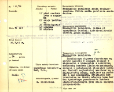
Title: File of histopathological evaluation of nervous system diseases (1966) - nr 112/66
Institutional creator:
Department of Experimental and Clinical Neuropathology MMRI
Contributor:
Description:
Clinical, anatomical and histological diagnosis
Abstract:
Histological diagnosis: Meningitis purulenta. Autopsy examination of 58-year-old patient was performed. Neuropathological evaluation in light microscopy was based on brain paraffin sections stained with Hematoxylin-eosin, cresyl-violet and van Gieson method.The main manifestation of the disease were abundant infiltrates in the meninges, composed mostly of leukocytes with a small admixture of lymphocyte-like cells, scavenger cells, and stimulated base elements. The infiltrates were most severe on the cerebral fornix, with minor infiltrates on the base and over the cerebellum. They generally did not progress to brain tissue. Very extensive injury was observed within the nerve cells, many of which showed features of ischemic disease. The cellular damage was more significant in the cerebral cortex than in the cerebellum, consistent with the severity of the inflammatory process in the adjacent meninges. In addition, there was massive blood stasis throughout the vascular system, both meningeal and interstitial, and in some areas perivascular transudates and petechiae were observed.
Format:
Resource Identifier:
Language:
Language of abstract:
Rights:
Creative Commons Attribution BY 4.0 license
Terms of use:
Copyright-protected material. [CC BY 4.0] May be used within the scope specified in Creative Commons Attribution BY 4.0 license, full text available at: ; -
Digitizing institution:
Mossakowski Medical Research Institute PAS
Original in:
Library of the Mossakowski Medical Research Institute PAS
Projects co-financed by:
Access:
Object collections:
- Digital Repository of Scientific Institutes > Scientific data and objects > Medical science > File of medical cases
- Digital Repository of Scientific Institutes > Partners' collections > Mossakowski Medical Research Institute PAS > Research data > File of histopathological evaluation of the nervous system diseases of the Department of Neuropathologu
Last modified:
Feb 1, 2022
In our library since:
Jun 22, 2021
Number of object content downloads / hits:
34
All available object's versions:
https://rcin.org.pl./publication/229338
Show description in RDF format:
Show description in RDFa format:
Show description in OAI-PMH format:
| Edition name | Date |
|---|---|
| opis nr 112/66 | Feb 1, 2022 |

 INSTYTUT ARCHEOLOGII I ETNOLOGII POLSKIEJ AKADEMII NAUK
 INSTYTUT BADAŃ LITERACKICH POLSKIEJ AKADEMII NAUK
 INSTYTUT BADAWCZY LEŚNICTWA
 INSTYTUT BIOLOGII DOŚWIADCZALNEJ IM. MARCELEGO NENCKIEGO POLSKIEJ AKADEMII NAUK
 INSTYTUT BIOLOGII SSAKÓW POLSKIEJ AKADEMII NAUK
 INSTYTUT CHEMII FIZYCZNEJ PAN
 INSTYTUT CHEMII ORGANICZNEJ PAN
 INSTYTUT FILOZOFII I SOCJOLOGII PAN
 INSTYTUT GEOGRAFII I PRZESTRZENNEGO ZAGOSPODAROWANIA PAN
 INSTYTUT HISTORII im. TADEUSZA MANTEUFFLA POLSKIEJ AKADEMII NAUK
 INSTYTUT JĘZYKA POLSKIEGO POLSKIEJ AKADEMII NAUK
 INSTYTUT MATEMATYCZNY PAN
 INSTYTUT MEDYCYNY DOŚWIADCZALNEJ I KLINICZNEJ IM.MIROSŁAWA MOSSAKOWSKIEGO POLSKIEJ AKADEMII NAUK
 INSTYTUT PODSTAWOWYCH PROBLEMÓW TECHNIKI PAN
 INSTYTUT SLAWISTYKI PAN
 SIEĆ BADAWCZA ŁUKASIEWICZ - INSTYTUT TECHNOLOGII MATERIAŁÓW ELEKTRONICZNYCH
 MUZEUM I INSTYTUT ZOOLOGII POLSKIEJ AKADEMII NAUK
 INSTYTUT BADAŃ SYSTEMOWYCH PAN
 INSTYTUT BOTANIKI IM. WŁADYSŁAWA SZAFERA POLSKIEJ AKADEMII NAUK


