- Search in all Repository
- Literature and maps
- Archeology
- Mills database
- Natural sciences
Advanced search
Advanced search
Advanced search
Advanced search
Advanced search

Object
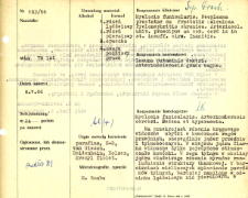
Title: Kartoteka oceny histopatologicznej chorób układu nerwowego (1966) - opis nr 103/66
Institutional creator:
Contributor:
H. Lewicka-Wysocka, lek. ; E. Osetowska, prof.
Publisher:
Description:
Rozpoznanie kliniczne, anatomiczne i histologiczne
Abstract:
Na przekrojach rdzenia kręgowego widoczne ubytki w komórkach rogów przednich, szczególnie jąder przednio i tylnobocznych. W miejscu jądra Clarka widoczne tylko pojedyncze stłuszczałe komórki. W rogu tylnym w okolicy jądra właściwego rdzenia wyraźne roz¬luźnienie struktury tkankowej. W bar¬wieniach mielinowych spłowienie powróź ków tylnych, bardziej nasilone w od¬cinku lędźwiowym niż piersiowym, z nieznaczną glejozą włóknistą, silniej wyrażoną w strukturach szarych.W korze opustoszenia komorkowe, zwyrodnienia tłuszczowe, schorzenia ischemiczne. W warstwie drobinowej zawoju hipokampa kule amyloidowe. W istocie białej zakrętu hipokampa między zakrętem zębatym a warstwą komórkową stopy hipokampa widoczna rozpływająca się prawie bezodczynowo tkanka.W całym OUN naczynia maksymalnie wypełnione krwią, ze ścianami zmienionymi szklisto i włóknisto. Przestrzenie Virchowa-Robina poszerzone Pogranicze tkankowo-naczyniowe rozluźnione, czasem przybiera charakter gąbczasty.W oponach wokół naczyń histiocyty, makrofagi i limfocyty.
Format:
Resource Identifier:
Language:
Language of abstract:
Rights:
Licencja Creative Commons Uznanie autorstwa 4.0
Terms of use:
Zasób chroniony prawem autorskim. [CC BY 4.0 Międzynarodowe] Korzystanie dozwolone zgodnie z licencją Creative Commons Uznanie autorstwa 4.0, której pełne postanowienia dostępne są pod adresem: ; -
Digitizing institution:
Instytut Medycyny Doświadczalnej i Klinicznej im. M. Mossakowskiego Polskiej Akademii Nauk
Original in:
Biblioteka Instytutu Medycyny Doświadczalnej i Klinicznej im. M. Mossakowskiego PAN
Projects co-financed by:
Access:
Object collections:
- Digital Repository of Scientific Institutes > Scientific data and objects > Medical science > File of medical cases
- Digital Repository of Scientific Institutes > Partners' collections > Mossakowski Medical Research Institute PAS > Research data > File of histopathological evaluation of the nervous system diseases of the Department of Neuropathologu
Last modified:
Feb 1, 2022
In our library since:
Jun 22, 2021
Number of object content downloads / hits:
49
All available object's versions:
https://rcin.org.pl./publication/229333
Show description in RDF format:
Show description in RDFa format:
Show description in OAI-PMH format:
| Edition name | Date |
|---|---|
| opis nr 103/66 | Feb 1, 2022 |
Objects Similar
Liberski, Paweł P Mossakowski, Mirosław Jan (1929–2001)
Grochowski, Witold

 INSTYTUT ARCHEOLOGII I ETNOLOGII POLSKIEJ AKADEMII NAUK
 INSTYTUT BADAŃ LITERACKICH POLSKIEJ AKADEMII NAUK
 INSTYTUT BADAWCZY LEŚNICTWA
 INSTYTUT BIOLOGII DOŚWIADCZALNEJ IM. MARCELEGO NENCKIEGO POLSKIEJ AKADEMII NAUK
 INSTYTUT BIOLOGII SSAKÓW POLSKIEJ AKADEMII NAUK
 INSTYTUT CHEMII FIZYCZNEJ PAN
 INSTYTUT CHEMII ORGANICZNEJ PAN
 INSTYTUT FILOZOFII I SOCJOLOGII PAN
 INSTYTUT GEOGRAFII I PRZESTRZENNEGO ZAGOSPODAROWANIA PAN
 INSTYTUT HISTORII im. TADEUSZA MANTEUFFLA POLSKIEJ AKADEMII NAUK
 INSTYTUT JĘZYKA POLSKIEGO POLSKIEJ AKADEMII NAUK
 INSTYTUT MATEMATYCZNY PAN
 INSTYTUT MEDYCYNY DOŚWIADCZALNEJ I KLINICZNEJ IM.MIROSŁAWA MOSSAKOWSKIEGO POLSKIEJ AKADEMII NAUK
 INSTYTUT PODSTAWOWYCH PROBLEMÓW TECHNIKI PAN
 INSTYTUT SLAWISTYKI PAN
 SIEĆ BADAWCZA ŁUKASIEWICZ - INSTYTUT TECHNOLOGII MATERIAŁÓW ELEKTRONICZNYCH
 MUZEUM I INSTYTUT ZOOLOGII POLSKIEJ AKADEMII NAUK
 INSTYTUT BADAŃ SYSTEMOWYCH PAN
 INSTYTUT BOTANIKI IM. WŁADYSŁAWA SZAFERA POLSKIEJ AKADEMII NAUK


