- Search in all Repository
- Literature and maps
- Archeology
- Mills database
- Natural sciences
Advanced search
Advanced search
Advanced search
Advanced search
Advanced search

Object
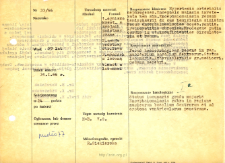
Title: File of histopathological evaluation of nervous system diseases (1966) - nr 33/66
Institutional creator:
Department of Experimental and Clinical Neuropathology MMRI
Contributor:
M. Marciniak ; K. Wiśniewska, dr ; E.Osetowska, prof.
Description:
Clinical, anatomical and histological diagnosis
Abstract:
Histological diagnosis: Encephalomalacia rubra. Autopsy examination of 59-year-old patient was performed. Neuropathological evaluation in light microscopy was based on brain paraffin sections stained with H-E and Van Gieson's method.Bilaterally, multiple foci of malacia in the dissection or glial-mesodermal organization stage were present in the basal ganglia, sometimes forming perivascular lacunae. Fresh blood was present in the ventricular system. Vascular endothelium was dilated. In the cerebellum, segmental atrophy of granular layer (Purkinje cells) was observed. In the white matter of the hemispheres an old focus of malacia with the formation of cavities was seen. Around the focus, altered astrocytes, paravascularly single macrophages and lymphocytes were seen.Fibrous and vitreous vascular changes, as well as features of congestion, edema, glial activation and proliferation were found throughout the nervous system. Fresh blood was present in the subarachnoid space.Histological diagnosis: Status lacunaris gradu majoris Encephalomalacia rubra in regione nucleorum basalium dextrorum et ad systema ventriculorum penetrans.
Format:
Resource Identifier:
Language:
Language of abstract:
Rights:
Creative Commons Attribution BY 4.0 license
Terms of use:
Copyright-protected material. [CC BY 4.0] May be used within the scope specified in Creative Commons Attribution BY 4.0 license, full text available at: ; -
Digitizing institution:
Mossakowski Medical Research Institute PAS
Original in:
Library of the Mossakowski Medical Research Institute PAS
Projects co-financed by:
Access:
Object collections:
- Digital Repository of Scientific Institutes > Scientific data and objects > Medical science > File of medical cases
- Digital Repository of Scientific Institutes > Partners' collections > Mossakowski Medical Research Institute PAS > Research data > File of histopathological evaluation of the nervous system diseases of the Department of Neuropathologu
Last modified:
Feb 1, 2022
In our library since:
Jun 21, 2021
Number of object content downloads / hits:
59
All available object's versions:
https://rcin.org.pl./publication/229191
Show description in RDF format:
Show description in RDFa format:
Show description in OAI-PMH format:
| Edition name | Date |
|---|---|
| opis nr 33/66 | Feb 1, 2022 |

 INSTYTUT ARCHEOLOGII I ETNOLOGII POLSKIEJ AKADEMII NAUK
 INSTYTUT BADAŃ LITERACKICH POLSKIEJ AKADEMII NAUK
 INSTYTUT BADAWCZY LEŚNICTWA
 INSTYTUT BIOLOGII DOŚWIADCZALNEJ IM. MARCELEGO NENCKIEGO POLSKIEJ AKADEMII NAUK
 INSTYTUT BIOLOGII SSAKÓW POLSKIEJ AKADEMII NAUK
 INSTYTUT CHEMII FIZYCZNEJ PAN
 INSTYTUT CHEMII ORGANICZNEJ PAN
 INSTYTUT FILOZOFII I SOCJOLOGII PAN
 INSTYTUT GEOGRAFII I PRZESTRZENNEGO ZAGOSPODAROWANIA PAN
 INSTYTUT HISTORII im. TADEUSZA MANTEUFFLA POLSKIEJ AKADEMII NAUK
 INSTYTUT JĘZYKA POLSKIEGO POLSKIEJ AKADEMII NAUK
 INSTYTUT MATEMATYCZNY PAN
 INSTYTUT MEDYCYNY DOŚWIADCZALNEJ I KLINICZNEJ IM.MIROSŁAWA MOSSAKOWSKIEGO POLSKIEJ AKADEMII NAUK
 INSTYTUT PODSTAWOWYCH PROBLEMÓW TECHNIKI PAN
 INSTYTUT SLAWISTYKI PAN
 SIEĆ BADAWCZA ŁUKASIEWICZ - INSTYTUT TECHNOLOGII MATERIAŁÓW ELEKTRONICZNYCH
 MUZEUM I INSTYTUT ZOOLOGII POLSKIEJ AKADEMII NAUK
 INSTYTUT BADAŃ SYSTEMOWYCH PAN
 INSTYTUT BOTANIKI IM. WŁADYSŁAWA SZAFERA POLSKIEJ AKADEMII NAUK


