- O repozytorium
- Kolekcje
- Indeksy
- Historia przeglądania
-
Repozytoria RCIN
-
 INSTYTUT ARCHEOLOGII I ETNOLOGII POLSKIEJ AKADEMII NAUK
-
 INSTYTUT BADAŃ LITERACKICH POLSKIEJ AKADEMII NAUK
-
 INSTYTUT BADAWCZY LEŚNICTWA
-
 INSTYTUT BIOLOGII DOŚWIADCZALNEJ IM. MARCELEGO NENCKIEGO POLSKIEJ AKADEMII NAUK
-
 INSTYTUT BIOLOGII SSAKÓW POLSKIEJ AKADEMII NAUK
-
 INSTYTUT CHEMII FIZYCZNEJ PAN
-
 INSTYTUT CHEMII ORGANICZNEJ PAN
-
 INSTYTUT FILOZOFII I SOCJOLOGII PAN
-
 INSTYTUT GEOGRAFII I PRZESTRZENNEGO ZAGOSPODAROWANIA PAN
-
 INSTYTUT HISTORII im. TADEUSZA MANTEUFFLA POLSKIEJ AKADEMII NAUK
-
 INSTYTUT JĘZYKA POLSKIEGO POLSKIEJ AKADEMII NAUK
-
 INSTYTUT MATEMATYCZNY PAN
-
 INSTYTUT MEDYCYNY DOŚWIADCZALNEJ I KLINICZNEJ IM.MIROSŁAWA MOSSAKOWSKIEGO POLSKIEJ AKADEMII NAUK
-
 INSTYTUT PODSTAWOWYCH PROBLEMÓW TECHNIKI PAN
-
 INSTYTUT SLAWISTYKI PAN
-
 SIEĆ BADAWCZA ŁUKASIEWICZ - INSTYTUT TECHNOLOGII MATERIAŁÓW ELEKTRONICZNYCH
-
 MUZEUM I INSTYTUT ZOOLOGII POLSKIEJ AKADEMII NAUK
-
 INSTYTUT BADAŃ SYSTEMOWYCH PAN
-
 INSTYTUT BOTANIKI IM. WŁADYSŁAWA SZAFERA POLSKIEJ AKADEMII NAUK
-
- Wyszukaj w całym Repozytorium
- Piśmiennictwo i mapy
- Archeologia
- Baza Młynów
- Nauki przyrodnicze
Wyszukiwanie zaawansowane
Wyszukiwanie zaawansowane
Wyszukiwanie zaawansowane
Wyszukiwanie zaawansowane
Wyszukiwanie zaawansowane

Obiekt
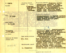
Tytuł: File of histopathological evaluation of nervous system diseases (1966) - nr 138/66
Twórca instytucjonalny:
Department of Experimental and Clinical Neuropathology MMRI
Współtwórca:
H. Lewicka-Wysocka, dr ; E. Osetowska, prof.
Opis:
Clinical, anatomical and histological diagnosis
Abstrakt:
Histological diagnosis: Encephalomalacia inveterata. Alba et partium rubra, in regione nucleorum basalium et insulae hemisphaerii dextri. Oedema et hyperaemia cerebri. Arteriosolerosis cerebri. Autopsy examination of 73-year-old patient was performed. Neuropathological evaluation in light microscopy was based on brain paraffin sections stained by H-E and Van Gieson's stain.In the basal ganglia a large focus of malacia, destroying the head of the caudate nucleus, the internal capsule and a part of the white matter of the frontal lobes was observed. In the centre a cavity in which remains of nervous tissue, mononuclear and multinuclear leucocytes, numerous partially haemolysed blood cells (sometimes containing hemosiderin), young vessels and collagen fibres were found was noticed. The second focus of malacia includes the cortex of the island. This area was in the dissection phase, macrophages, rod-shaped microglia, stimulated vessels and glia were visible. In the cortex and basal ganglia, chronic, ischemic and fatty degeneration was observed. Tissue was loosen at the border of this focus. Vessels filled with blood and significant atherosclerotic lesions of vitreous-fibrous type in vessel walls were seen. In the meninges, around the vessels single lymphocytes, histiocytes were present.
Format:
Identyfikator zasobu:
Język:
Język streszczenia:
Prawa:
Creative Commons Attribution BY 4.0 license
Zasady wykorzystania:
Copyright-protected material. [CC BY 4.0] May be used within the scope specified in Creative Commons Attribution BY 4.0 license, full text available at: ; -
Digitalizacja:
Mossakowski Medical Research Institute PAS
Lokalizacja oryginału:
Library of the Mossakowski Medical Research Institute PAS
Dofinansowane ze środków:
Dostęp:
Kolekcje, do których przypisany jest obiekt:
- Repozytorium Cyfrowe Instytutów Naukowych > Dane i obiekty naukowe > Nauki medyczne > Kartoteka przypadków medycznych
- Repozytorium Cyfrowe Instytutów Naukowych > Kolekcje Partnerów > Instytut Medycyny Doświadczalnej i Klinicznej PAN > Dane Badawcze > Kartoteka oceny histopatologicznej chorób układu nerwowego Zakładu Neuropatologii
Data ostatniej modyfikacji:
1 lut 2022
Data dodania obiektu:
22 cze 2021
Liczba pobrań / odtworzeń:
49
Wszystkie dostępne wersje tego obiektu:
https://rcin.org.pl./publication/229355
Wyświetl opis w formacie RDF:
Wyświetl opis w formacie RDFa:
Wyświetl opis w formacie OAI-PMH:
| Nazwa wydania | Data |
|---|---|
| opis nr 138/66 | 1 lut 2022 |


