- About project
- Collections
- Indexes
- Recently viewed
-
RCIN Repositories
-
 INSTYTUT ARCHEOLOGII I ETNOLOGII POLSKIEJ AKADEMII NAUK
-
 INSTYTUT BADAŃ LITERACKICH POLSKIEJ AKADEMII NAUK
-
 INSTYTUT BADAWCZY LEŚNICTWA
-
 INSTYTUT BIOLOGII DOŚWIADCZALNEJ IM. MARCELEGO NENCKIEGO POLSKIEJ AKADEMII NAUK
-
 INSTYTUT BIOLOGII SSAKÓW POLSKIEJ AKADEMII NAUK
-
 INSTYTUT CHEMII FIZYCZNEJ PAN
-
 INSTYTUT CHEMII ORGANICZNEJ PAN
-
 INSTYTUT FILOZOFII I SOCJOLOGII PAN
-
 INSTYTUT GEOGRAFII I PRZESTRZENNEGO ZAGOSPODAROWANIA PAN
-
 INSTYTUT HISTORII im. TADEUSZA MANTEUFFLA POLSKIEJ AKADEMII NAUK
-
 INSTYTUT JĘZYKA POLSKIEGO POLSKIEJ AKADEMII NAUK
-
 INSTYTUT MATEMATYCZNY PAN
-
 INSTYTUT MEDYCYNY DOŚWIADCZALNEJ I KLINICZNEJ IM.MIROSŁAWA MOSSAKOWSKIEGO POLSKIEJ AKADEMII NAUK
-
 INSTYTUT PODSTAWOWYCH PROBLEMÓW TECHNIKI PAN
-
 INSTYTUT SLAWISTYKI PAN
-
 SIEĆ BADAWCZA ŁUKASIEWICZ - INSTYTUT TECHNOLOGII MATERIAŁÓW ELEKTRONICZNYCH
-
 MUZEUM I INSTYTUT ZOOLOGII POLSKIEJ AKADEMII NAUK
-
 INSTYTUT BADAŃ SYSTEMOWYCH PAN
-
 INSTYTUT BOTANIKI IM. WŁADYSŁAWA SZAFERA POLSKIEJ AKADEMII NAUK
-
- Search in all Repository
- Literature and maps
- Archeology
- Mills database
- Natural sciences
Advanced search
Advanced search
Advanced search
Advanced search
Advanced search

Object
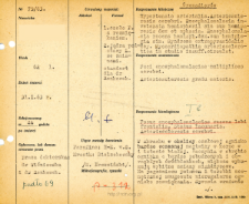
Title: File of histopathological evaluation of nervous system diseases (1963) - nr 79/63
Institutional creator:
Department of Experimental and Clinical Neuropathology MMRI
Contributor:
Place of publishing:
Description:
Clinical, anatomical and histological diagnosis
Abstract:
Histological diagnosis: Focus encephalomalaciae recens lobi frontalis, Stus lacunaris.Autopsy examination of 62-year-old patient was performed. Neuropathological evaluation in light microscopy was based on brain sections stained with Hematoxylin-eosin and van Gieson and brain frozen sections stained with Bielschowsky method.A focus of very early necrosis was found in the frontal area, in the cortex and white matter. The vessels within the focus were characterized by highly stimulated and swollen endothelium, and larger vessels showed prominent atherosclerotic changes. An irregular focus of malacia was seen at the border of the putamen and capsula externa. Some perivascular areas became extensive sinuses and only peripherally more or less abundant clusters of macrophages were observed, sometimes with admixture of histiocytes and single fibroblasts. In the vicinity of this focus, a distinct density of cellular glia was seen. The vessels had atheromatically altered walls /atheromatous, vitreous, fibrous-vitreous/ with symptomatic lymphocytic infiltrates. Accurate assessment of the extent of cellular damage and edema was impossible due to decay.
Format:
Resource Identifier:
Language:
Language of abstract:
Rights:
Creative Commons Attribution BY 4.0 license
Terms of use:
Copyright-protected material. [CC BY 4.0] May be used within the scope specified in Creative Commons Attribution BY 4.0 license, full text available at: ; -
Digitizing institution:
Mossakowski Medical Research Institute PAS
Original in:
Library of the Mossakowski Medical Research Institute PAS
Projects co-financed by:
Access:
Object collections:
- Digital Repository of Scientific Institutes > Scientific data and objects > Medical science > File of medical cases
- Digital Repository of Scientific Institutes > Partners' collections > Mossakowski Medical Research Institute PAS > Research data > File of histopathological evaluation of the nervous system diseases of the Department of Neuropathologu
Last modified:
Feb 2, 2022
In our library since:
Sep 10, 2021
Number of object content downloads / hits:
44
All available object's versions:
https://rcin.org.pl./publication/246244
Show description in RDF format:
Show description in RDFa format:
Show description in OAI-PMH format:
| Edition name | Date |
|---|---|
| opis nr 79/63 | Feb 2, 2022 |


