- About project
- Collections
- Indexes
- Recently viewed
-
RCIN Repositories
-
 INSTYTUT ARCHEOLOGII I ETNOLOGII POLSKIEJ AKADEMII NAUK
-
 INSTYTUT BADAŃ LITERACKICH POLSKIEJ AKADEMII NAUK
-
 INSTYTUT BADAWCZY LEŚNICTWA
-
 INSTYTUT BIOLOGII DOŚWIADCZALNEJ IM. MARCELEGO NENCKIEGO POLSKIEJ AKADEMII NAUK
-
 INSTYTUT BIOLOGII SSAKÓW POLSKIEJ AKADEMII NAUK
-
 INSTYTUT CHEMII FIZYCZNEJ PAN
-
 INSTYTUT CHEMII ORGANICZNEJ PAN
-
 INSTYTUT FILOZOFII I SOCJOLOGII PAN
-
 INSTYTUT GEOGRAFII I PRZESTRZENNEGO ZAGOSPODAROWANIA PAN
-
 INSTYTUT HISTORII im. TADEUSZA MANTEUFFLA POLSKIEJ AKADEMII NAUK
-
 INSTYTUT JĘZYKA POLSKIEGO POLSKIEJ AKADEMII NAUK
-
 INSTYTUT MATEMATYCZNY PAN
-
 INSTYTUT MEDYCYNY DOŚWIADCZALNEJ I KLINICZNEJ IM.MIROSŁAWA MOSSAKOWSKIEGO POLSKIEJ AKADEMII NAUK
-
 INSTYTUT PODSTAWOWYCH PROBLEMÓW TECHNIKI PAN
-
 INSTYTUT SLAWISTYKI PAN
-
 SIEĆ BADAWCZA ŁUKASIEWICZ - INSTYTUT TECHNOLOGII MATERIAŁÓW ELEKTRONICZNYCH
-
 MUZEUM I INSTYTUT ZOOLOGII POLSKIEJ AKADEMII NAUK
-
 INSTYTUT BADAŃ SYSTEMOWYCH PAN
-
 INSTYTUT BOTANIKI IM. WŁADYSŁAWA SZAFERA POLSKIEJ AKADEMII NAUK
-
- Search in all Repository
- Literature and maps
- Archeology
- Mills database
- Natural sciences
Advanced search
Advanced search
Advanced search
Advanced search
Advanced search

Object
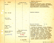
Title: File of histopathological evaluation of nervous system diseases (1965) - nr 80/65
Institutional creator:
Department of Experimental and Clinical Neuropathology MMRI
Contributor:
Place of publishing:
Description:
Clinical, anatomical and histological diagnosis
Abstract:
Histological diagnosis: Meningitis purulenta. Autopsy examination of 53-year-old patient was performed. Neuropathological evaluation in light microscopy was based on brain paraffin sections stained with Hematoxylin-eosin, cresyl-violet and Van Gieson's method.Fibrous exudate with leukocytic infiltration was found in the meninges at the top of the left frontal lobe superior ganglion. Besides, infiltrates of plasma cells, histio or lymphocyte-like cells were present scattered loosely in the connective tissue and perivascularly in the meninges and cerebellum. Plasma cells were sometimes multinucleated. Here and there inflammatory infiltration from meninges penetrated deep into the brain tissue, often through V.R. spaces. A spongiosis-like state was observed, as well as the formation of microabscesses. In the cortex, cellular atrophy, ischemic disorders and steatosis were evident. There were gemistiocytes in the fine layer of the cortex. In the cerebellum, steatosis of the dentate nucleus and Bergman's glial proliferation were observed. Besides, features of congestion, edema, proliferation and glial activation were found throughout the nervous system.
Format:
Resource Identifier:
Language:
Language of abstract:
Rights:
Creative Commons Attribution BY 4.0 license
Terms of use:
Copyright-protected material. [CC BY 4.0] May be used within the scope specified in Creative Commons Attribution BY 4.0 license, full text available at: ; -
Digitizing institution:
Mossakowski Medical Research Institute PAS
Original in:
Library of the Mossakowski Medical Research Institute PAS
Projects co-financed by:
Access:
Object collections:
- Digital Repository of Scientific Institutes > Scientific data and objects > Medical science > File of medical cases
- Digital Repository of Scientific Institutes > Partners' collections > Mossakowski Medical Research Institute PAS > Research data > File of histopathological evaluation of the nervous system diseases of the Department of Neuropathologu
Last modified:
Feb 1, 2022
In our library since:
Jul 9, 2021
Number of object content downloads / hits:
44
All available object's versions:
https://rcin.org.pl./publication/232822
Show description in RDF format:
Show description in RDFa format:
Show description in OAI-PMH format:
| Edition name | Date |
|---|---|
| opis nr 80/65 | Feb 1, 2022 |


