- About project
- Collections
- Indexes
- Recently viewed
-
RCIN Repositories
-
 INSTYTUT ARCHEOLOGII I ETNOLOGII POLSKIEJ AKADEMII NAUK
-
 INSTYTUT BADAŃ LITERACKICH POLSKIEJ AKADEMII NAUK
-
 INSTYTUT BADAWCZY LEŚNICTWA
-
 INSTYTUT BIOLOGII DOŚWIADCZALNEJ IM. MARCELEGO NENCKIEGO POLSKIEJ AKADEMII NAUK
-
 INSTYTUT BIOLOGII SSAKÓW POLSKIEJ AKADEMII NAUK
-
 INSTYTUT CHEMII FIZYCZNEJ PAN
-
 INSTYTUT CHEMII ORGANICZNEJ PAN
-
 INSTYTUT FILOZOFII I SOCJOLOGII PAN
-
 INSTYTUT GEOGRAFII I PRZESTRZENNEGO ZAGOSPODAROWANIA PAN
-
 INSTYTUT HISTORII im. TADEUSZA MANTEUFFLA POLSKIEJ AKADEMII NAUK
-
 INSTYTUT JĘZYKA POLSKIEGO POLSKIEJ AKADEMII NAUK
-
 INSTYTUT MATEMATYCZNY PAN
-
 INSTYTUT MEDYCYNY DOŚWIADCZALNEJ I KLINICZNEJ IM.MIROSŁAWA MOSSAKOWSKIEGO POLSKIEJ AKADEMII NAUK
-
 INSTYTUT PODSTAWOWYCH PROBLEMÓW TECHNIKI PAN
-
 INSTYTUT SLAWISTYKI PAN
-
 SIEĆ BADAWCZA ŁUKASIEWICZ - INSTYTUT TECHNOLOGII MATERIAŁÓW ELEKTRONICZNYCH
-
 MUZEUM I INSTYTUT ZOOLOGII POLSKIEJ AKADEMII NAUK
-
 INSTYTUT BADAŃ SYSTEMOWYCH PAN
-
 INSTYTUT BOTANIKI IM. WŁADYSŁAWA SZAFERA POLSKIEJ AKADEMII NAUK
-
- Search in all Repository
- Literature and maps
- Archeology
- Mills database
- Natural sciences
Advanced search
Advanced search
Advanced search
Advanced search
Advanced search

Object
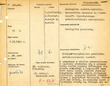
Title: File of histopathological evaluation of nervous system diseases (1962) - nr 231/62
Institutional creator:
Department of Experimental and Clinical Neuropathology MMRI
Contributor:
Place of publishing:
Description:
Clinical, anatomical and histological diagnosis
Abstract:
Autopsy examination of 73-year-old patient was performed. Neuropathological evaluation in light microscopy was based on brain paraffin sections stained with Hematoxylin-eosin and van Gieson method.In specimens from the prefrontal and frontal regions, a very abundant inflammatory infiltrate was found in the meninges, consisting primarily of leukocytes and a fairly large number of mononuclear cells, among which lymphocytes, occasionally plasma cells, and single histocytes were seen. More sparse inflammatory lesions were observed in the partially preserved meninges of the pons. The infiltrates penetrated deep into the cortex, along the venous vessels, sometimes also into the superficial layers of the cortex. Vascular inflammatory infiltrates were more abundant in the cortex than in the white matter of the cerebral hemispheres in the pons, mostly affecting the subcortical area. The infiltrates were composed mainly of mononuclear cells and did not extend beyond the vessel wall, and often together with edematous endothelium they led to the closure of the lumen of small veins without any obvious reaction from the surrounding tissue. There were very advanced vitreous lesions in the vasa vasorum. In the brain tissue, the fibro-vitreous changes were somewhat less pronounced. In the frontal and prefrontal cortex, large fields of cellular metastasis were seen, and in the cells, ischemic changes and fatty degeneration were seen. Diffuse oligodendroglial thickening was observed in the white matter of the cerebral hemispheres. Swelling in the white matter with widening of the V-R space and sponge-like thinning of the vascular fiber tissue was prominent.Histological diagnosis: Meningo-encephalitis purulenta. Arteriosclerosis cerebri.
Format:
Resource Identifier:
Language:
Language of abstract:
Rights:
Creative Commons Attribution BY 4.0 license
Terms of use:
Copyright-protected material. [CC BY 4.0] May be used within the scope specified in Creative Commons Attribution BY 4.0 license, full text available at: ; -
Digitizing institution:
Mossakowski Medical Research Institute PAS
Original in:
Library of the Mossakowski Medical Research Institute PAS
Projects co-financed by:
Access:
Object collections:
- Digital Repository of Scientific Institutes > Scientific data and objects > Medical science > File of medical cases
- Digital Repository of Scientific Institutes > Partners' collections > Mossakowski Medical Research Institute PAS > Research data > File of histopathological evaluation of the nervous system diseases of the Department of Neuropathologu
Last modified:
Mar 29, 2022
In our library since:
Oct 1, 2021
Number of object content downloads / hits:
83
All available object's versions:
https://rcin.org.pl./publication/251121
Show description in RDF format:
Show description in RDFa format:
Show description in OAI-PMH format:
| Edition name | Date |
|---|---|
| opis nr 231/62 | Mar 29, 2022 |


