- Wyszukaj w całym Repozytorium
- Piśmiennictwo i mapy
- Archeologia
- Baza Młynów
- Nauki przyrodnicze
Wyszukiwanie zaawansowane
Wyszukiwanie zaawansowane
Wyszukiwanie zaawansowane
Wyszukiwanie zaawansowane
Wyszukiwanie zaawansowane

Obiekt
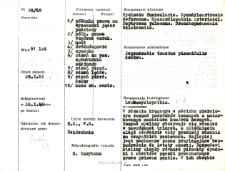
Tytuł: Kartoteka oceny histopatologicznej chorób układu nerwowego (1965) - opis nr 28/65
Twórca instytucjonalny:
Współtwórca:
Miejsce wydania:
Opis:
Rozpoznanie kliniczne, anatomiczne i histologiczne
Abstrakt:
W rdzeniu kręgowym w odcinku piersiowym rozpad powrózków bocznych z umiarkowanym naciekiem komórek żernych. Rozpad myeliny obserwuje się równieżw powrózkach tylnych, a zblednięciu uległ właściwie cały przekrój rdzeniana wszystkich poziomach. Najlepiej jest zachowane pasmo przylegające bezpośrednio do istoty szarej. Rozpadowi mieliny uległy również piramidy opuszki i niektóre pęczki przechodzące przez griseum pontis. W ich obrębie występują pojedyncze komórki żerne. W rdzeniu piersiowym w istocie białej nacieki okołonaczyniowe drobno okrągło komórkowe,W całym rdzeniu uogólniona glejoza, Komórki nerwowe obfitują w lipofuscyne, W przekrojach przez półkule prawą móźgu uogólnione zgąbczenie kory, istoty białej i jąder podstawy /chyba artefakt/ oraz skąpe naciekiw poszerzonych przestrzeniach okołonaczyniowych, Naczynia: niektóre drobnetętnice i tętniczki ze zmianami szklistymi.Obraz morfologiczny mógłby odpowiadać myelosis funicularis, gdyby stan kliniczny za tą jednostką nozologiczną przemawiał. Dane kliniczne o dostępnym skrócie historii choroby /brak zmian we krwi obwodowej, brak rozmazu szpiku i bite zaburzeń czucia/ nie pozwalają na pewne rozpoznanie zwyrodnienia powrózkowatego.
Format:
Identyfikator zasobu:
Język:
Język streszczenia:
Prawa:
Licencja Creative Commons Uznanie autorstwa 4.0
Zasady wykorzystania:
Zasób chroniony prawem autorskim. [CC BY 4.0 Międzynarodowe] Korzystanie dozwolone zgodnie z licencją Creative Commons Uznanie autorstwa 4.0, której pełne postanowienia dostępne są pod adresem: ; -
Digitalizacja:
Instytut Medycyny Doświadczalnej i Klinicznej im. M. Mossakowskiego Polskiej Akademii Nauk
Lokalizacja oryginału:
Biblioteka Instytutu Medycyny Doświadczalnej i Klinicznej im. M. Mossakowskiego PAN
Dofinansowane ze środków:
Dostęp:
Kolekcje, do których przypisany jest obiekt:
- Repozytorium Cyfrowe Instytutów Naukowych > Dane i obiekty naukowe > Nauki medyczne > Kartoteka przypadków medycznych
- Repozytorium Cyfrowe Instytutów Naukowych > Kolekcje Partnerów > Instytut Medycyny Doświadczalnej i Klinicznej PAN > Dane Badawcze > Kartoteka oceny histopatologicznej chorób układu nerwowego Zakładu Neuropatologii
Data ostatniej modyfikacji:
1 lut 2022
Data dodania obiektu:
8 lip 2021
Liczba pobrań / odtworzeń:
53
Wszystkie dostępne wersje tego obiektu:
https://rcin.org.pl./publication/232779
Wyświetl opis w formacie RDF:
Wyświetl opis w formacie RDFa:
Wyświetl opis w formacie OAI-PMH:
| Nazwa wydania | Data |
|---|---|
| opis nr 28/65 | 1 lut 2022 |

 INSTYTUT ARCHEOLOGII I ETNOLOGII POLSKIEJ AKADEMII NAUK
 INSTYTUT BADAŃ LITERACKICH POLSKIEJ AKADEMII NAUK
 INSTYTUT BADAWCZY LEŚNICTWA
 INSTYTUT BIOLOGII DOŚWIADCZALNEJ IM. MARCELEGO NENCKIEGO POLSKIEJ AKADEMII NAUK
 INSTYTUT BIOLOGII SSAKÓW POLSKIEJ AKADEMII NAUK
 INSTYTUT CHEMII FIZYCZNEJ PAN
 INSTYTUT CHEMII ORGANICZNEJ PAN
 INSTYTUT FILOZOFII I SOCJOLOGII PAN
 INSTYTUT GEOGRAFII I PRZESTRZENNEGO ZAGOSPODAROWANIA PAN
 INSTYTUT HISTORII im. TADEUSZA MANTEUFFLA POLSKIEJ AKADEMII NAUK
 INSTYTUT JĘZYKA POLSKIEGO POLSKIEJ AKADEMII NAUK
 INSTYTUT MATEMATYCZNY PAN
 INSTYTUT MEDYCYNY DOŚWIADCZALNEJ I KLINICZNEJ IM.MIROSŁAWA MOSSAKOWSKIEGO POLSKIEJ AKADEMII NAUK
 INSTYTUT PODSTAWOWYCH PROBLEMÓW TECHNIKI PAN
 INSTYTUT SLAWISTYKI PAN
 SIEĆ BADAWCZA ŁUKASIEWICZ - INSTYTUT TECHNOLOGII MATERIAŁÓW ELEKTRONICZNYCH
 MUZEUM I INSTYTUT ZOOLOGII POLSKIEJ AKADEMII NAUK
 INSTYTUT BADAŃ SYSTEMOWYCH PAN
 INSTYTUT BOTANIKI IM. WŁADYSŁAWA SZAFERA POLSKIEJ AKADEMII NAUK


