- O repozytorium
- Kolekcje
- Indeksy
- Historia przeglądania
-
Repozytoria RCIN
-
 INSTYTUT ARCHEOLOGII I ETNOLOGII POLSKIEJ AKADEMII NAUK
-
 INSTYTUT BADAŃ LITERACKICH POLSKIEJ AKADEMII NAUK
-
 INSTYTUT BADAWCZY LEŚNICTWA
-
 INSTYTUT BIOLOGII DOŚWIADCZALNEJ IM. MARCELEGO NENCKIEGO POLSKIEJ AKADEMII NAUK
-
 INSTYTUT BIOLOGII SSAKÓW POLSKIEJ AKADEMII NAUK
-
 INSTYTUT CHEMII FIZYCZNEJ PAN
-
 INSTYTUT CHEMII ORGANICZNEJ PAN
-
 INSTYTUT FILOZOFII I SOCJOLOGII PAN
-
 INSTYTUT GEOGRAFII I PRZESTRZENNEGO ZAGOSPODAROWANIA PAN
-
 INSTYTUT HISTORII im. TADEUSZA MANTEUFFLA POLSKIEJ AKADEMII NAUK
-
 INSTYTUT JĘZYKA POLSKIEGO POLSKIEJ AKADEMII NAUK
-
 INSTYTUT MATEMATYCZNY PAN
-
 INSTYTUT MEDYCYNY DOŚWIADCZALNEJ I KLINICZNEJ IM.MIROSŁAWA MOSSAKOWSKIEGO POLSKIEJ AKADEMII NAUK
-
 INSTYTUT PODSTAWOWYCH PROBLEMÓW TECHNIKI PAN
-
 INSTYTUT SLAWISTYKI PAN
-
 SIEĆ BADAWCZA ŁUKASIEWICZ - INSTYTUT TECHNOLOGII MATERIAŁÓW ELEKTRONICZNYCH
-
 MUZEUM I INSTYTUT ZOOLOGII POLSKIEJ AKADEMII NAUK
-
 INSTYTUT BADAŃ SYSTEMOWYCH PAN
-
 INSTYTUT BOTANIKI IM. WŁADYSŁAWA SZAFERA POLSKIEJ AKADEMII NAUK
-
- Wyszukaj w całym Repozytorium
- Piśmiennictwo i mapy
- Archeologia
- Baza Młynów
- Nauki przyrodnicze
Wyszukiwanie zaawansowane
Wyszukiwanie zaawansowane
Wyszukiwanie zaawansowane
Wyszukiwanie zaawansowane
Wyszukiwanie zaawansowane

Obiekt
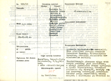
Tytuł: File of histopathological evaluation of nervous system diseases (1965) - nr 119/65
Institutional creator:
Department of Experimental and Clinical Neuropathology MMRI
Contributor:
Place of publishing:
Description:
Clinical, anatomical and histological diagnosis
Abstract:
Histological diagnosis: Agenesis hemisphaerium cerebelli. Vere similiter atresia completa foraminum Zuschka. Hydrocephalus internus. Atrophia secundaria hemisphaerium cerebri.Autopsy examination of 2,5-year-old patient was performed. Neuropathological evaluation in light microscopy was based on brain paraffin sections stained with Cresyl-violet, Heidenhain, Holzer, Bojan and Van Gieson's method.The structure of the cerebral mantle and basal ganglia was normal and the gyrus, although small, were present in adequate number. However, a huge, bloated ventricular system was found, which probably compressed the brain tissue and pushed it peripherally towards the cranial bones. Due to this compression, the circulation was impaired. Arterial and venous vessels had thick, fibrotic walls and were surrounded by "lakes" of the transudate. Neuronal atrophy was observed in the cortex and basal nuclei and compensatory proliferation of cellular and fibrous glia. The meninges were thick, and numerous fresh extravasations were found in them, as well as evidence of old hemorrhages in the form of hemosiderin. The cause of all these abnormalities appears to be malformation of the cerebellum. The cerebellum developed only in the central part, the cerebellar hemispheres were completely missing. Enormous internal hydrocephalus pushed ventricle IV, which surrounded almost all around the brainstem structures, expanded ventricle III and lateral ventricles, causing an image of secondary cerebral atrophy. Vascular changes and epidural hemorrhages should be interpreted as secondary. There were no data enabling the diagnosis of primary inflammatory process or post-necrotic scarring. Therefore, the picture should be considered as a result of congenital malformation of the cerebellum and its connections with the brainstem.Histological diagnosis: Agenesis hemisphaerium cerebelli. Vere similiter atresia completa foraminum Zuschka. Hydrocephalus internus. Atrophia secundaria hemisphaerium cerebri.Autopsy examination of 2,5-year-old patient was performed. Neuropathological evaluation in light microscopy was based on brain paraffin sections stained with Cresyl-violet, Heidenhain, Holzer, Bojan and Van Gieson's method.The structure of the cerebral mantle and basal ganglia was normal and the gyrus, although small, were present in adequate number. However, a huge, bloated ventricular system was found, which probably compressed the brain tissue and pushed it peripherally towards the cranial bones. Due to this compression, the circulation was impaired. Arterial and venous vessels had thick, fibrotic walls and were surrounded by "lakes" of the transudate. Neuronal atrophy was observed in the cortex and basal nuclei and compensatory proliferation of cellular and fibrous glia. The meninges were thick, and numerous fresh extravasations were found in them, as well as evidence of old hemorrhages in the form of hemosiderin. The cause of all these abnormalities appears to be malformation of the cerebellum. The cerebellum developed only in the central part, the cerebellar hemispheres were completely missing. Enormous internal hydrocephalus pushed ventricle IV, which surrounded almost all around the brainstem structures, expanded ventricle III and lateral ventricles, causing an image of secondary cerebral atrophy. Vascular changes and epidural hemorrhages should be interpreted as secondary. There were no data enabling the diagnosis of primary inflammatory process or post-necrotic scarring. Therefore, the picture should be considered as a result of congenital malformation of the cerebellum and its connections with the brainstem.Histological diagnosis: Agenesis hemisphaerium cerebelli. Vere similiter atresia completa foraminum Zuschka. Hydrocephalus internus. Atrophia secundaria hemisphaerium cerebri.
Format:
Resource Identifier:
Language:
Language of abstract:
Rights:
Creative Commons Attribution BY 4.0 license
Terms of use:
Copyright-protected material. [CC BY 4.0] May be used within the scope specified in Creative Commons Attribution BY 4.0 license, full text available at: ; -
Digitizing institution:
Mossakowski Medical Research Institute PAS
Original in:
Library of the Mossakowski Medical Research Institute PAS
Projects co-financed by:
Access:
Kolekcje, do których przypisany jest obiekt:
- Digital Repository of Scientific Institutes > Scientific data and objects > Medical science > File of medical cases
- Digital Repository of Scientific Institutes > Partners' collections > Mossakowski Medical Research Institute PAS > Research data > File of histopathological evaluation of the nervous system diseases of the Department of Neuropathologu
Data ostatniej modyfikacji:
Feb 1, 2022
Data dodania obiektu:
Jul 9, 2021
Liczba pobrań / odtworzeń:
58
Wszystkie dostępne wersje tego obiektu:
https://rcin.org.pl./publication/232845
Wyświetl opis w formacie RDF:
Wyświetl opis w formacie RDFa:
Wyświetl opis w formacie OAI-PMH:
| Nazwa wydania | Data |
|---|---|
| opis nr 119/65 | Feb 1, 2022 |
Obiekty Podobne
Mossakowski, Mirosław Jan (1929–2001)


