- O repozytorium
- Kolekcje
- Indeksy
- Historia przeglądania
-
Repozytoria RCIN
-
 INSTYTUT ARCHEOLOGII I ETNOLOGII POLSKIEJ AKADEMII NAUK
-
 INSTYTUT BADAŃ LITERACKICH POLSKIEJ AKADEMII NAUK
-
 INSTYTUT BADAWCZY LEŚNICTWA
-
 INSTYTUT BIOLOGII DOŚWIADCZALNEJ IM. MARCELEGO NENCKIEGO POLSKIEJ AKADEMII NAUK
-
 INSTYTUT BIOLOGII SSAKÓW POLSKIEJ AKADEMII NAUK
-
 INSTYTUT CHEMII FIZYCZNEJ PAN
-
 INSTYTUT CHEMII ORGANICZNEJ PAN
-
 INSTYTUT FILOZOFII I SOCJOLOGII PAN
-
 INSTYTUT GEOGRAFII I PRZESTRZENNEGO ZAGOSPODAROWANIA PAN
-
 INSTYTUT HISTORII im. TADEUSZA MANTEUFFLA POLSKIEJ AKADEMII NAUK
-
 INSTYTUT JĘZYKA POLSKIEGO POLSKIEJ AKADEMII NAUK
-
 INSTYTUT MATEMATYCZNY PAN
-
 INSTYTUT MEDYCYNY DOŚWIADCZALNEJ I KLINICZNEJ IM.MIROSŁAWA MOSSAKOWSKIEGO POLSKIEJ AKADEMII NAUK
-
 INSTYTUT PODSTAWOWYCH PROBLEMÓW TECHNIKI PAN
-
 INSTYTUT SLAWISTYKI PAN
-
 SIEĆ BADAWCZA ŁUKASIEWICZ - INSTYTUT TECHNOLOGII MATERIAŁÓW ELEKTRONICZNYCH
-
 MUZEUM I INSTYTUT ZOOLOGII POLSKIEJ AKADEMII NAUK
-
 INSTYTUT BADAŃ SYSTEMOWYCH PAN
-
 INSTYTUT BOTANIKI IM. WŁADYSŁAWA SZAFERA POLSKIEJ AKADEMII NAUK
-
- Wyszukaj w całym Repozytorium
- Piśmiennictwo i mapy
- Archeologia
- Baza Młynów
- Nauki przyrodnicze
Wyszukiwanie zaawansowane
Wyszukiwanie zaawansowane
Wyszukiwanie zaawansowane
Wyszukiwanie zaawansowane
Wyszukiwanie zaawansowane

Obiekt
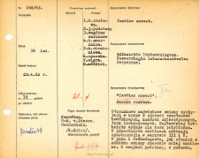
Tytuł: File of histopathological evaluation of nervous system diseases (1963) - nr 168/63
Twórca instytucjonalny:
Department of Experimental and Clinical Neuropathology MMRI
Współtwórca:
Miejsce wydania:
Opis:
Clinical, anatomical and histological diagnosis
Abstrakt:
Histological diagnosis: ”Cardiac arrest”. Anoxia cerebri.Autopsy examination of 56-year-old patient was performed. Neuropathological evaluation in light microscopy was based on brain paraffin sections stained with Hematoxylin-eosin, Heidenhain and van Gieson method.The relatively most severe lesions were in the cortex in the form of cell vacuolation, sclerotic cells, and ischemic forms. The occipital cortex was the most affected. In other areas, vacuolations were found in layers III and V. In the brainstem and midbrain, as well as in the cerebellum, the lesions were much less severe. In the basal ganglia, in the putamen, tissue thinning was found at the small and medium vessels. The thalamus nuclei were relatively well preserved. There was no glial reaction. In the vessels, fairly advanced sclerotic lesions were observed.
Format:
Identyfikator zasobu:
Język:
Język streszczenia:
Prawa:
Creative Commons Attribution BY 4.0 license
Zasady wykorzystania:
Copyright-protected material. [CC BY 4.0] May be used within the scope specified in Creative Commons Attribution BY 4.0 license, full text available at: ; -
Digitalizacja:
Mossakowski Medical Research Institute PAS
Lokalizacja oryginału:
Library of the Mossakowski Medical Research Institute PAS
Dofinansowane ze środków:
Dostęp:
Kolekcje, do których przypisany jest obiekt:
- Repozytorium Cyfrowe Instytutów Naukowych > Dane i obiekty naukowe > Nauki medyczne > Kartoteka przypadków medycznych
- Repozytorium Cyfrowe Instytutów Naukowych > Kolekcje Partnerów > Instytut Medycyny Doświadczalnej i Klinicznej PAN > Dane Badawcze > Kartoteka oceny histopatologicznej chorób układu nerwowego Zakładu Neuropatologii
Data ostatniej modyfikacji:
1 lut 2022
Data dodania obiektu:
14 wrz 2021
Liczba pobrań / odtworzeń:
73
Wszystkie dostępne wersje tego obiektu:
https://rcin.org.pl./publication/246307
Wyświetl opis w formacie RDF:
Wyświetl opis w formacie RDFa:
Wyświetl opis w formacie OAI-PMH:
| Nazwa wydania | Data |
|---|---|
| opis nr 168/63 | 1 lut 2022 |


