- Search in all Repository
- Literature and maps
- Archeology
- Mills database
- Natural sciences
Advanced search
Advanced search
Advanced search
Advanced search
Advanced search

Object
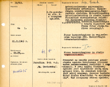
Title: File of histopathological evaluation of nervous system diseases (1963) - nr 32/63
Institutional creator:
Department of Experimental and Clinical Neuropathology MMRI
Contributor:
Place of publishing:
Description:
Clinical, anatomical and histological diagnosis
Abstract:
Histological diagnosis: Focus haemorrhagicus in stadio organisationis.Autopsy examination of 64-year-old patient was performed. Neuropathological evaluation in light microscopy was based on brain paraffin sections stained with Hematoxylin-eosin and van Gieson.A hemorrhagic focus was found in the basal ganglia. In the central part, the focus was filled with a solid mass of blood; toward the periphery, the hemorrhage was less massive. A reaction was visible around the focus, containing a network of proliferating capillaries and fibroblasts that penetrated into the focus. Very peripherally, glial activation was evident. Near the ventricle, in a small section, the reaction did not directly surround the hemorrhagic focus; a field was found there that looked as if blood was pressing the necrotic tissue. Overall, the microscopic image indicates an age of the focus consistent with the duration of the disease, but necrotic changes to the tissue into which the hemorrhage occurred cannot be excluded.
Format:
Resource Identifier:
Language:
Language of abstract:
Rights:
Creative Commons Attribution BY 4.0 license
Terms of use:
Copyright-protected material. [CC BY 4.0] May be used within the scope specified in Creative Commons Attribution BY 4.0 license, full text available at: ; -
Digitizing institution:
Mossakowski Medical Research Institute PAS
Original in:
Library of the Mossakowski Medical Research Institute PAS
Projects co-financed by:
Access:
Object collections:
- Digital Repository of Scientific Institutes > Scientific data and objects > Medical science > File of medical cases
- Digital Repository of Scientific Institutes > Partners' collections > Mossakowski Medical Research Institute PAS > Research data > File of histopathological evaluation of the nervous system diseases of the Department of Neuropathologu
Last modified:
Feb 1, 2022
In our library since:
Sep 10, 2021
Number of object content downloads / hits:
40
All available object's versions:
https://rcin.org.pl./publication/246232
Show description in RDF format:
Show description in RDFa format:
Show description in OAI-PMH format:
| Edition name | Date |
|---|---|
| opis nr 32/63 | Feb 1, 2022 |

 INSTYTUT ARCHEOLOGII I ETNOLOGII POLSKIEJ AKADEMII NAUK
 INSTYTUT BADAŃ LITERACKICH POLSKIEJ AKADEMII NAUK
 INSTYTUT BADAWCZY LEŚNICTWA
 INSTYTUT BIOLOGII DOŚWIADCZALNEJ IM. MARCELEGO NENCKIEGO POLSKIEJ AKADEMII NAUK
 INSTYTUT BIOLOGII SSAKÓW POLSKIEJ AKADEMII NAUK
 INSTYTUT CHEMII FIZYCZNEJ PAN
 INSTYTUT CHEMII ORGANICZNEJ PAN
 INSTYTUT FILOZOFII I SOCJOLOGII PAN
 INSTYTUT GEOGRAFII I PRZESTRZENNEGO ZAGOSPODAROWANIA PAN
 INSTYTUT HISTORII im. TADEUSZA MANTEUFFLA POLSKIEJ AKADEMII NAUK
 INSTYTUT JĘZYKA POLSKIEGO POLSKIEJ AKADEMII NAUK
 INSTYTUT MATEMATYCZNY PAN
 INSTYTUT MEDYCYNY DOŚWIADCZALNEJ I KLINICZNEJ IM.MIROSŁAWA MOSSAKOWSKIEGO POLSKIEJ AKADEMII NAUK
 INSTYTUT PODSTAWOWYCH PROBLEMÓW TECHNIKI PAN
 INSTYTUT SLAWISTYKI PAN
 SIEĆ BADAWCZA ŁUKASIEWICZ - INSTYTUT TECHNOLOGII MATERIAŁÓW ELEKTRONICZNYCH
 MUZEUM I INSTYTUT ZOOLOGII POLSKIEJ AKADEMII NAUK
 INSTYTUT BADAŃ SYSTEMOWYCH PAN
 INSTYTUT BOTANIKI IM. WŁADYSŁAWA SZAFERA POLSKIEJ AKADEMII NAUK


