- O repozytorium
- Kolekcje
- Indeksy
- Historia przeglądania
-
Repozytoria RCIN
-
 INSTYTUT ARCHEOLOGII I ETNOLOGII POLSKIEJ AKADEMII NAUK
-
 INSTYTUT BADAŃ LITERACKICH POLSKIEJ AKADEMII NAUK
-
 INSTYTUT BADAWCZY LEŚNICTWA
-
 INSTYTUT BIOLOGII DOŚWIADCZALNEJ IM. MARCELEGO NENCKIEGO POLSKIEJ AKADEMII NAUK
-
 INSTYTUT BIOLOGII SSAKÓW POLSKIEJ AKADEMII NAUK
-
 INSTYTUT CHEMII FIZYCZNEJ PAN
-
 INSTYTUT CHEMII ORGANICZNEJ PAN
-
 INSTYTUT FILOZOFII I SOCJOLOGII PAN
-
 INSTYTUT GEOGRAFII I PRZESTRZENNEGO ZAGOSPODAROWANIA PAN
-
 INSTYTUT HISTORII im. TADEUSZA MANTEUFFLA POLSKIEJ AKADEMII NAUK
-
 INSTYTUT JĘZYKA POLSKIEGO POLSKIEJ AKADEMII NAUK
-
 INSTYTUT MATEMATYCZNY PAN
-
 INSTYTUT MEDYCYNY DOŚWIADCZALNEJ I KLINICZNEJ IM.MIROSŁAWA MOSSAKOWSKIEGO POLSKIEJ AKADEMII NAUK
-
 INSTYTUT PODSTAWOWYCH PROBLEMÓW TECHNIKI PAN
-
 INSTYTUT SLAWISTYKI PAN
-
 SIEĆ BADAWCZA ŁUKASIEWICZ - INSTYTUT TECHNOLOGII MATERIAŁÓW ELEKTRONICZNYCH
-
 MUZEUM I INSTYTUT ZOOLOGII POLSKIEJ AKADEMII NAUK
-
 INSTYTUT BADAŃ SYSTEMOWYCH PAN
-
 INSTYTUT BOTANIKI IM. WŁADYSŁAWA SZAFERA POLSKIEJ AKADEMII NAUK
-
- Wyszukaj w całym Repozytorium
- Piśmiennictwo i mapy
- Archeologia
- Baza Młynów
- Nauki przyrodnicze
Wyszukiwanie zaawansowane
Wyszukiwanie zaawansowane
Wyszukiwanie zaawansowane
Wyszukiwanie zaawansowane
Wyszukiwanie zaawansowane

Obiekt
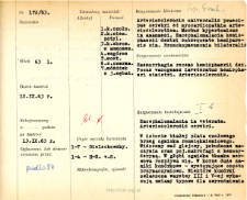
Tytuł: Kartoteka oceny histopatologicznej chorób układu nerwowego (1963) - opis nr 172/63
Twórca instytucjonalny:
Współtwórca:
Miejsce wydania:
Opis:
Rozpoznanie kliniczne, anatomiczne i histologiczne
Abstrakt:
W istocie białej płata czołowegobrzeg ogniska rozmięknieniowego.Widoczny wał glejowy, pobudzone naczynia oraz poj.makrofagi z hemosyderyną. W głębi ogniska tkanka nerwowa rozpływa się. W korze duże zaniki komórkowe - występujące często przynaczyniowo. Niektóre komórki zwłaszcza warstwy III i V-ej wykazują zmiany typowe dla zwyrodnienia tłuszczowego, w niektórych widoczna tigroliza. Duże zmiany zwyrodnienia tłuszczowego obserwuje się w komórkach, wzgórza i w jądrze zębatym. Naczynia wykazują duże zmiany stwardnieniowe /szkliste i włókniste/. Najmniejsze zmiany naczyniowe widoczne są w korze /tylko włókniste/. Poza tym cechy przekrwienia. W oponach gdzieniegdzie pojedyncze komórki histocytarne,makrofagi.
Format:
Identyfikator zasobu:
Język:
Język streszczenia:
Prawa:
Licencja Creative Commons Uznanie autorstwa 4.0
Zasady wykorzystania:
Zasób chroniony prawem autorskim. [CC BY 4.0 Międzynarodowe] Korzystanie dozwolone zgodnie z licencją Creative Commons Uznanie autorstwa 4.0, której pełne postanowienia dostępne są pod adresem: ; -
Digitalizacja:
Instytut Medycyny Doświadczalnej i Klinicznej im. M. Mossakowskiego Polskiej Akademii Nauk
Lokalizacja oryginału:
Biblioteka Instytutu Medycyny Doświadczalnej i Klinicznej im. M. Mossakowskiego PAN
Dofinansowane ze środków:
Dostęp:
Kolekcje, do których przypisany jest obiekt:
- Repozytorium Cyfrowe Instytutów Naukowych > Dane i obiekty naukowe > Nauki medyczne > Kartoteka przypadków medycznych
- Repozytorium Cyfrowe Instytutów Naukowych > Kolekcje Partnerów > Instytut Medycyny Doświadczalnej i Klinicznej PAN > Dane Badawcze > Kartoteka oceny histopatologicznej chorób układu nerwowego Zakładu Neuropatologii
Data ostatniej modyfikacji:
1 lut 2022
Data dodania obiektu:
14 wrz 2021
Liczba pobrań / odtworzeń:
60
Wszystkie dostępne wersje tego obiektu:
https://rcin.org.pl./publication/246308
Wyświetl opis w formacie RDF:
Wyświetl opis w formacie RDFa:
Wyświetl opis w formacie OAI-PMH:
| Nazwa wydania | Data |
|---|---|
| opis nr 172/63 | 1 lut 2022 |


