- Search in all Repository
- Literature and maps
- Archeology
- Mills database
- Natural sciences
Advanced search
Advanced search
Advanced search
Advanced search
Advanced search

Object
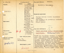
Title: File of histopathological evaluation of nervous system diseases (1962) - nr 154/62
Institutional creator:
Department of Experimental and Clinical Neuropathology MMRI
Contributor:
Place of publishing:
Description:
Clinical, anatomical and histological diagnosis
Abstract:
Histological diagnosis: Tumor medullae spin glioblastoma multiforme. Sarkomatosis.Autopsy examination of patient was performed. Neuropathological evaluation in light microscopy was based on brain paraffin sections stained with Hematoxylin-eosin, van Gieson and Heidenhain method.The medulla in the sections below and above the tumor was swollen and compressed; the structure of the grey matter was distorted, the horns pushed far apart. The cells of the anterior horns were considerably thinned, and showed far-reaching tigrolysis or even images of homogenous disease. The tumor had an exceedingly abundant cellular composition. It contained polymorphic cells: round, spindle-shaped, rod-shaped; many of them resembled mast cell astrocytes. There were extremely many two- and three-nucleated cells, as well as giant cells with a small number of hyperchromatic nuclei. There were also nuclear conglomerates without visible protoplasm. The tumor contained numerous mitoses (several in the field of view) and often images of amitosis were visible. In the vicinity of the tumor the proliferation of the sheath elements was observed, the sheath cells were stimulated, large, proliferating.
Format:
Resource Identifier:
Language:
Language of abstract:
Rights:
Creative Commons Attribution BY 4.0 license
Terms of use:
Copyright-protected material. [CC BY 4.0] May be used within the scope specified in Creative Commons Attribution BY 4.0 license, full text available at: ; -
Digitizing institution:
Mossakowski Medical Research Institute PAS
Original in:
Library of the Mossakowski Medical Research Institute PAS
Projects co-financed by:
Access:
Object collections:
- Digital Repository of Scientific Institutes > Scientific data and objects > Medical science > File of medical cases
- Digital Repository of Scientific Institutes > Partners' collections > Mossakowski Medical Research Institute PAS > Research data > File of histopathological evaluation of the nervous system diseases of the Department of Neuropathologu
Last modified:
Feb 1, 2022
In our library since:
Oct 1, 2021
Number of object content downloads / hits:
73
All available object's versions:
https://rcin.org.pl./publication/251068
Show description in RDF format:
Show description in RDFa format:
Show description in OAI-PMH format:
| Edition name | Date |
|---|---|
| opis nr 154/62 | Feb 1, 2022 |

 INSTYTUT ARCHEOLOGII I ETNOLOGII POLSKIEJ AKADEMII NAUK
 INSTYTUT BADAŃ LITERACKICH POLSKIEJ AKADEMII NAUK
 INSTYTUT BADAWCZY LEŚNICTWA
 INSTYTUT BIOLOGII DOŚWIADCZALNEJ IM. MARCELEGO NENCKIEGO POLSKIEJ AKADEMII NAUK
 INSTYTUT BIOLOGII SSAKÓW POLSKIEJ AKADEMII NAUK
 INSTYTUT CHEMII FIZYCZNEJ PAN
 INSTYTUT CHEMII ORGANICZNEJ PAN
 INSTYTUT FILOZOFII I SOCJOLOGII PAN
 INSTYTUT GEOGRAFII I PRZESTRZENNEGO ZAGOSPODAROWANIA PAN
 INSTYTUT HISTORII im. TADEUSZA MANTEUFFLA POLSKIEJ AKADEMII NAUK
 INSTYTUT JĘZYKA POLSKIEGO POLSKIEJ AKADEMII NAUK
 INSTYTUT MATEMATYCZNY PAN
 INSTYTUT MEDYCYNY DOŚWIADCZALNEJ I KLINICZNEJ IM.MIROSŁAWA MOSSAKOWSKIEGO POLSKIEJ AKADEMII NAUK
 INSTYTUT PODSTAWOWYCH PROBLEMÓW TECHNIKI PAN
 INSTYTUT SLAWISTYKI PAN
 SIEĆ BADAWCZA ŁUKASIEWICZ - INSTYTUT TECHNOLOGII MATERIAŁÓW ELEKTRONICZNYCH
 MUZEUM I INSTYTUT ZOOLOGII POLSKIEJ AKADEMII NAUK
 INSTYTUT BADAŃ SYSTEMOWYCH PAN
 INSTYTUT BOTANIKI IM. WŁADYSŁAWA SZAFERA POLSKIEJ AKADEMII NAUK


