- O repozytorium
- Kolekcje
- Indeksy
- Historia przeglądania
-
Repozytoria RCIN
-
 INSTYTUT ARCHEOLOGII I ETNOLOGII POLSKIEJ AKADEMII NAUK
-
 INSTYTUT BADAŃ LITERACKICH POLSKIEJ AKADEMII NAUK
-
 INSTYTUT BADAWCZY LEŚNICTWA
-
 INSTYTUT BIOLOGII DOŚWIADCZALNEJ IM. MARCELEGO NENCKIEGO POLSKIEJ AKADEMII NAUK
-
 INSTYTUT BIOLOGII SSAKÓW POLSKIEJ AKADEMII NAUK
-
 INSTYTUT CHEMII FIZYCZNEJ PAN
-
 INSTYTUT CHEMII ORGANICZNEJ PAN
-
 INSTYTUT FILOZOFII I SOCJOLOGII PAN
-
 INSTYTUT GEOGRAFII I PRZESTRZENNEGO ZAGOSPODAROWANIA PAN
-
 INSTYTUT HISTORII im. TADEUSZA MANTEUFFLA POLSKIEJ AKADEMII NAUK
-
 INSTYTUT JĘZYKA POLSKIEGO POLSKIEJ AKADEMII NAUK
-
 INSTYTUT MATEMATYCZNY PAN
-
 INSTYTUT MEDYCYNY DOŚWIADCZALNEJ I KLINICZNEJ IM.MIROSŁAWA MOSSAKOWSKIEGO POLSKIEJ AKADEMII NAUK
-
 INSTYTUT PODSTAWOWYCH PROBLEMÓW TECHNIKI PAN
-
 INSTYTUT SLAWISTYKI PAN
-
 SIEĆ BADAWCZA ŁUKASIEWICZ - INSTYTUT TECHNOLOGII MATERIAŁÓW ELEKTRONICZNYCH
-
 MUZEUM I INSTYTUT ZOOLOGII POLSKIEJ AKADEMII NAUK
-
 INSTYTUT BADAŃ SYSTEMOWYCH PAN
-
 INSTYTUT BOTANIKI IM. WŁADYSŁAWA SZAFERA POLSKIEJ AKADEMII NAUK
-

Obiekt
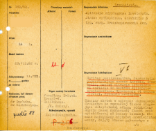
Tytuł: File of histopathological evaluation of nervous system diseases (1962) - nr 161/62
Twórca instytucjonalny:
Department of Experimental and Clinical Neuropathology MMRI
Współtwórca:
Miejsce wydania:
Opis:
Clinical, anatomical and histological diagnosis
Abstrakt:
Autopsy examination of 24-year-old patient was performed. Neuropathological evaluation in light microscopy was based on brain paraffin sections stained with Hematoxylin-eosin, van Gieson, cresyl - violet and Heidenhain method.A large cavity after tissue disintegration was found in the medial temporal gyrus. The area most closely resembled the defect - demyelinated, covered by a spongy lesion with thinning of the glia, almost exclusively astrocytes remained. The edge of the cavity was hypertrophied with connective tissue fibers, with occasional single macrophages visible in the cavity lumen. The hippocampal gyrus showed numerous cellular defects especially in the Sommer sector. The white matter of this gyrus was partially covered by spongiosis. Nerve cells in almost all specimens showed alterations involving tigrolysis, severe cellular or ischemic changes. Most Purkinje cells were homogenously altered. In addition to Sommer's sector, uneven cellular loss of inferior olive, Purkinje cells and numerous cortical vacuolations were found. The vessels were fibrotic, most filled with blood. The adventitial spaces were almost always dilated, showing sparse round cell elements and single macrophages with hemosiderin. The nerve fibers of the white matter were dispersed. In locally, small free spaces of transudate fluid could be seen. A doubtful 2-nucleus nerve cell was visible in the globus pallidus. The meninges were thickened and fibrotic.Histological diagnosis: Cavitas posttraumatica. Degeneratio parenchymatosa cornus Ammonis, cerebelli, nuclei olivaris inferioris at corticis in decursu epilepsiae symptomaticae. Hyperaemia et oedema cerebri. Fibrosis meningum.
Format:
Identyfikator zasobu:
Język:
Język streszczenia:
Prawa:
Creative Commons Attribution BY 4.0 license
Zasady wykorzystania:
Copyright-protected material. [CC BY 4.0] May be used within the scope specified in Creative Commons Attribution BY 4.0 license, full text available at: ; -
Digitalizacja:
Mossakowski Medical Research Institute PAS
Lokalizacja oryginału:
Library of the Mossakowski Medical Research Institute PAS
Dofinansowane ze środków:
Dostęp:
Kolekcje, do których przypisany jest obiekt:
- Repozytorium Cyfrowe Instytutów Naukowych > Dane i obiekty naukowe > Nauki medyczne > Kartoteka przypadków medycznych
- Repozytorium Cyfrowe Instytutów Naukowych > Kolekcje Partnerów > Instytut Medycyny Doświadczalnej i Klinicznej PAN > Dane Badawcze > Kartoteka oceny histopatologicznej chorób układu nerwowego Zakładu Neuropatologii
Data ostatniej modyfikacji:
29 mar 2022
Data dodania obiektu:
1 paź 2021
Liczba pobrań / odtworzeń:
59
Wszystkie dostępne wersje tego obiektu:
https://rcin.org.pl./publication/251071
Wyświetl opis w formacie RDF:
Wyświetl opis w formacie RDFa:
Wyświetl opis w formacie OAI-PMH:
| Nazwa wydania | Data |
|---|---|
| opis nr 161/62 | 29 mar 2022 |


