- O repozytorium
- Kolekcje
- Indeksy
- Historia przeglądania
-
Repozytoria RCIN
-
 INSTYTUT ARCHEOLOGII I ETNOLOGII POLSKIEJ AKADEMII NAUK
-
 INSTYTUT BADAŃ LITERACKICH POLSKIEJ AKADEMII NAUK
-
 INSTYTUT BADAWCZY LEŚNICTWA
-
 INSTYTUT BIOLOGII DOŚWIADCZALNEJ IM. MARCELEGO NENCKIEGO POLSKIEJ AKADEMII NAUK
-
 INSTYTUT BIOLOGII SSAKÓW POLSKIEJ AKADEMII NAUK
-
 INSTYTUT CHEMII FIZYCZNEJ PAN
-
 INSTYTUT CHEMII ORGANICZNEJ PAN
-
 INSTYTUT FILOZOFII I SOCJOLOGII PAN
-
 INSTYTUT GEOGRAFII I PRZESTRZENNEGO ZAGOSPODAROWANIA PAN
-
 INSTYTUT HISTORII im. TADEUSZA MANTEUFFLA POLSKIEJ AKADEMII NAUK
-
 INSTYTUT JĘZYKA POLSKIEGO POLSKIEJ AKADEMII NAUK
-
 INSTYTUT MATEMATYCZNY PAN
-
 INSTYTUT MEDYCYNY DOŚWIADCZALNEJ I KLINICZNEJ IM.MIROSŁAWA MOSSAKOWSKIEGO POLSKIEJ AKADEMII NAUK
-
 INSTYTUT PODSTAWOWYCH PROBLEMÓW TECHNIKI PAN
-
 INSTYTUT SLAWISTYKI PAN
-
 SIEĆ BADAWCZA ŁUKASIEWICZ - INSTYTUT TECHNOLOGII MATERIAŁÓW ELEKTRONICZNYCH
-
 MUZEUM I INSTYTUT ZOOLOGII POLSKIEJ AKADEMII NAUK
-
 INSTYTUT BADAŃ SYSTEMOWYCH PAN
-
 INSTYTUT BOTANIKI IM. WŁADYSŁAWA SZAFERA POLSKIEJ AKADEMII NAUK
-

Obiekt
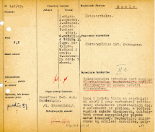
Tytuł: File of histopathological evaluation of nervous system diseases (1963) - nr 147/63
Twórca instytucjonalny:
Department of Experimental and Clinical Neuropathology MMRI
Współtwórca:
Miejsce wydania:
Opis:
Clinical, anatomical and histological diagnosis
Abstrakt:
Histological diagnosis: Hydrocephalus internus post meningo ependimitidem. Haemorrhagia subarachnoidealis et oedema cerebri secundarium. Autopsy examination of 5,5-year-old patient was performed. Neuropathological evaluation in light microscopy was based on brain paraffin sections stained with Hematoxylin-eosin, Heidenhain and van Gieson method.The cortex and white matter were pushed to the periphery by the fluid-expanded ventricular system. Moving from the meninges towards the ventricular system, the following changes were found, which were more or less the same at all levels: the meninges were edematous, in some areas extremely thick, with a rich proliferation of collagen fibers. Remnant lymphocytic infiltrates were seen among the fibers and perivascularly. In addition, fresh subarachnoid extravasations were seen, locally taking the form of extensive hemorrhages. The frontal, right occipital cortex was formed normally, but with large cellular atrophy. The white matter was edematous, loosened in structure, with prominent astrocyte proliferation and significant stasis of capillaries and medium-sized vessels. The most characteristic lesions were found in the subependymal area of the temporal horn: several vessels with lymphocytic-plasmacytic infiltrates were found there, as well as two clusters of granulomatous infiltrate passing to the ventricular walls. Along the entire ventricular system, spongiotic necrosis with proliferation of mast cell and fibrous glia was seen. The picture was the evidence of an inflammatory process, most likely a simple purulent bacterial case, which had migrated from the basal meninges to the ependyma, leading to progressive closure of the ventricular system. Subsequently, the inflammatory case healed /the type of infiltration indicates this/, and the hydrocephalus, increasing with scarring, led to a descent with symptoms of edema and hemorrhage.
Format:
Identyfikator zasobu:
Język:
Język streszczenia:
Prawa:
Creative Commons Attribution BY 4.0 license
Zasady wykorzystania:
Copyright-protected material. [CC BY 4.0] May be used within the scope specified in Creative Commons Attribution BY 4.0 license, full text available at: ; -
Digitalizacja:
Mossakowski Medical Research Institute PAS
Lokalizacja oryginału:
Library of the Mossakowski Medical Research Institute PAS
Dofinansowane ze środków:
Dostęp:
Kolekcje, do których przypisany jest obiekt:
- Repozytorium Cyfrowe Instytutów Naukowych > Dane i obiekty naukowe > Nauki medyczne > Kartoteka przypadków medycznych
- Repozytorium Cyfrowe Instytutów Naukowych > Kolekcje Partnerów > Instytut Medycyny Doświadczalnej i Klinicznej PAN > Dane Badawcze > Kartoteka oceny histopatologicznej chorób układu nerwowego Zakładu Neuropatologii
Data ostatniej modyfikacji:
1 lut 2022
Data dodania obiektu:
14 wrz 2021
Liczba pobrań / odtworzeń:
36
Wszystkie dostępne wersje tego obiektu:
https://rcin.org.pl./publication/246299
Wyświetl opis w formacie RDF:
Wyświetl opis w formacie RDFa:
Wyświetl opis w formacie OAI-PMH:
| Nazwa wydania | Data |
|---|---|
| opis nr 147/63 | 1 lut 2022 |


