- O repozytorium
- Kolekcje
- Indeksy
- Historia przeglądania
-
Repozytoria RCIN
-
 INSTYTUT ARCHEOLOGII I ETNOLOGII POLSKIEJ AKADEMII NAUK
-
 INSTYTUT BADAŃ LITERACKICH POLSKIEJ AKADEMII NAUK
-
 INSTYTUT BADAWCZY LEŚNICTWA
-
 INSTYTUT BIOLOGII DOŚWIADCZALNEJ IM. MARCELEGO NENCKIEGO POLSKIEJ AKADEMII NAUK
-
 INSTYTUT BIOLOGII SSAKÓW POLSKIEJ AKADEMII NAUK
-
 INSTYTUT CHEMII FIZYCZNEJ PAN
-
 INSTYTUT CHEMII ORGANICZNEJ PAN
-
 INSTYTUT FILOZOFII I SOCJOLOGII PAN
-
 INSTYTUT GEOGRAFII I PRZESTRZENNEGO ZAGOSPODAROWANIA PAN
-
 INSTYTUT HISTORII im. TADEUSZA MANTEUFFLA POLSKIEJ AKADEMII NAUK
-
 INSTYTUT JĘZYKA POLSKIEGO POLSKIEJ AKADEMII NAUK
-
 INSTYTUT MATEMATYCZNY PAN
-
 INSTYTUT MEDYCYNY DOŚWIADCZALNEJ I KLINICZNEJ IM.MIROSŁAWA MOSSAKOWSKIEGO POLSKIEJ AKADEMII NAUK
-
 INSTYTUT PODSTAWOWYCH PROBLEMÓW TECHNIKI PAN
-
 INSTYTUT SLAWISTYKI PAN
-
 SIEĆ BADAWCZA ŁUKASIEWICZ - INSTYTUT TECHNOLOGII MATERIAŁÓW ELEKTRONICZNYCH
-
 MUZEUM I INSTYTUT ZOOLOGII POLSKIEJ AKADEMII NAUK
-
 INSTYTUT BADAŃ SYSTEMOWYCH PAN
-
 INSTYTUT BOTANIKI IM. WŁADYSŁAWA SZAFERA POLSKIEJ AKADEMII NAUK
-
- Wyszukaj w całym Repozytorium
- Piśmiennictwo i mapy
- Archeologia
- Baza Młynów
- Nauki przyrodnicze
Wyszukiwanie zaawansowane
Wyszukiwanie zaawansowane
Wyszukiwanie zaawansowane
Wyszukiwanie zaawansowane
Wyszukiwanie zaawansowane

Obiekt
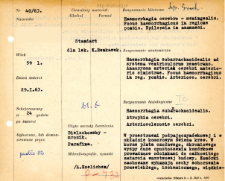
Tytuł: File of histopathological evaluation of nervous system diseases (1963) - nr 40/63
Twórca instytucjonalny:
Department of Experimental and Clinical Neuropathology MMRI
Współtwórca:
Miejsce wydania:
Opis:
Clinical, anatomical and histological diagnosis
Abstrakt:
Histological diagnosis: Haemorrhagia subarachnoidealis. Atrophia cerebri. Arteriosclerosis cerebri.Autopsy examination of 59-year-old patient was performed. Neuropathological evaluation in light microscopy was based on brain frozen sections stained with Bielschowsky method.Fresh blood was present in the subarachnoid space and ventricular system. In the cortex of the frontal, temporal, and insular lobes, large cellular vacuolations were evident, leading in sections to complete obliteration of the layered structure. Preserved cells showed features of chronic, ischemic, severe disease; some were steatotic. Blood was found in the globus pallidus around some vessels. The vessels were fibrously altered. Besides, features of congestion, glial activation and proliferation were observed. Single Alzheimer's fibers were visualized in Bielschowsky staining. No senile plaques were found.
Format:
Identyfikator zasobu:
Język:
Język streszczenia:
Prawa:
Creative Commons Attribution BY 4.0 license
Zasady wykorzystania:
Copyright-protected material. [CC BY 4.0] May be used within the scope specified in Creative Commons Attribution BY 4.0 license, full text available at: ; -
Digitalizacja:
Mossakowski Medical Research Institute PAS
Lokalizacja oryginału:
Library of the Mossakowski Medical Research Institute PAS
Dofinansowane ze środków:
Dostęp:
Kolekcje, do których przypisany jest obiekt:
- Repozytorium Cyfrowe Instytutów Naukowych > Dane i obiekty naukowe > Nauki medyczne > Kartoteka przypadków medycznych
- Repozytorium Cyfrowe Instytutów Naukowych > Kolekcje Partnerów > Instytut Medycyny Doświadczalnej i Klinicznej PAN > Dane Badawcze > Kartoteka oceny histopatologicznej chorób układu nerwowego Zakładu Neuropatologii
Data ostatniej modyfikacji:
2 lut 2022
Data dodania obiektu:
10 wrz 2021
Liczba pobrań / odtworzeń:
50
Wszystkie dostępne wersje tego obiektu:
https://rcin.org.pl./publication/246239
Wyświetl opis w formacie RDF:
Wyświetl opis w formacie RDFa:
Wyświetl opis w formacie OAI-PMH:
| Nazwa wydania | Data |
|---|---|
| opis nr 40/63 | 2 lut 2022 |


