- About project
- Collections
- Indexes
- Recently viewed
-
RCIN Repositories
-
 INSTYTUT ARCHEOLOGII I ETNOLOGII POLSKIEJ AKADEMII NAUK
-
 INSTYTUT BADAŃ LITERACKICH POLSKIEJ AKADEMII NAUK
-
 INSTYTUT BADAWCZY LEŚNICTWA
-
 INSTYTUT BIOLOGII DOŚWIADCZALNEJ IM. MARCELEGO NENCKIEGO POLSKIEJ AKADEMII NAUK
-
 INSTYTUT BIOLOGII SSAKÓW POLSKIEJ AKADEMII NAUK
-
 INSTYTUT CHEMII FIZYCZNEJ PAN
-
 INSTYTUT CHEMII ORGANICZNEJ PAN
-
 INSTYTUT FILOZOFII I SOCJOLOGII PAN
-
 INSTYTUT GEOGRAFII I PRZESTRZENNEGO ZAGOSPODAROWANIA PAN
-
 INSTYTUT HISTORII im. TADEUSZA MANTEUFFLA POLSKIEJ AKADEMII NAUK
-
 INSTYTUT JĘZYKA POLSKIEGO POLSKIEJ AKADEMII NAUK
-
 INSTYTUT MATEMATYCZNY PAN
-
 INSTYTUT MEDYCYNY DOŚWIADCZALNEJ I KLINICZNEJ IM.MIROSŁAWA MOSSAKOWSKIEGO POLSKIEJ AKADEMII NAUK
-
 INSTYTUT PODSTAWOWYCH PROBLEMÓW TECHNIKI PAN
-
 INSTYTUT SLAWISTYKI PAN
-
 SIEĆ BADAWCZA ŁUKASIEWICZ - INSTYTUT TECHNOLOGII MATERIAŁÓW ELEKTRONICZNYCH
-
 MUZEUM I INSTYTUT ZOOLOGII POLSKIEJ AKADEMII NAUK
-
 INSTYTUT BADAŃ SYSTEMOWYCH PAN
-
 INSTYTUT BOTANIKI IM. WŁADYSŁAWA SZAFERA POLSKIEJ AKADEMII NAUK
-
- Search in all Repository
- Literature and maps
- Archeology
- Mills database
- Natural sciences
Advanced search
Advanced search
Advanced search
Advanced search
Advanced search

Object
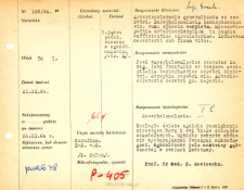
Title: File of histopathological evaluation of nervous system diseases (1964) - nr 188/64
Institutional creator:
Department of Experimental and Clinical Neuropathology MMRI
Contributor:
Place of publishing:
Description:
Clinical, anatomical and histological diagnosis
Abstract:
Histological diagnosis: Encephalomalacia.Autopsy examination of 78-year-old patient was performed. Neuropathological evaluation in light microscopy was based on brain paraffin sections stained with Hematoxylin-eosin and van Gieson method.Extensive fresh foci of malacia were found, destroying the cortex and white axes of the orbital gyri. The foci were covered with abundant fresh macrophages. Limited fields of spongy necrosis were present in the basal ganglia
Format:
Resource Identifier:
Language:
Language of abstract:
Rights:
Creative Commons Attribution BY 4.0 license
Terms of use:
Copyright-protected material. [CC BY 4.0] May be used within the scope specified in Creative Commons Attribution BY 4.0 license, full text available at: ; -
Digitizing institution:
Mossakowski Medical Research Institute PAS
Original in:
Library of the Mossakowski Medical Research Institute PAS
Projects co-financed by:
Access:
Object collections:
- Digital Repository of Scientific Institutes > Scientific data and objects > Medical science > File of medical cases
- Digital Repository of Scientific Institutes > Partners' collections > Mossakowski Medical Research Institute PAS > Research data > File of histopathological evaluation of the nervous system diseases of the Department of Neuropathologu
Last modified:
Oct 11, 2022
In our library since:
Aug 20, 2021
Number of object content downloads / hits:
37
All available object's versions:
https://rcin.org.pl./publication/241488
Show description in RDF format:
Show description in RDFa format:
Show description in OAI-PMH format:
| Edition name | Date |
|---|---|
| opis nr 188/64 | Oct 11, 2022 |


