- About project
- Collections
- Indexes
- Recently viewed
-
RCIN Repositories
-
 INSTYTUT ARCHEOLOGII I ETNOLOGII POLSKIEJ AKADEMII NAUK
-
 INSTYTUT BADAŃ LITERACKICH POLSKIEJ AKADEMII NAUK
-
 INSTYTUT BADAWCZY LEŚNICTWA
-
 INSTYTUT BIOLOGII DOŚWIADCZALNEJ IM. MARCELEGO NENCKIEGO POLSKIEJ AKADEMII NAUK
-
 INSTYTUT BIOLOGII SSAKÓW POLSKIEJ AKADEMII NAUK
-
 INSTYTUT CHEMII FIZYCZNEJ PAN
-
 INSTYTUT CHEMII ORGANICZNEJ PAN
-
 INSTYTUT FILOZOFII I SOCJOLOGII PAN
-
 INSTYTUT GEOGRAFII I PRZESTRZENNEGO ZAGOSPODAROWANIA PAN
-
 INSTYTUT HISTORII im. TADEUSZA MANTEUFFLA POLSKIEJ AKADEMII NAUK
-
 INSTYTUT JĘZYKA POLSKIEGO POLSKIEJ AKADEMII NAUK
-
 INSTYTUT MATEMATYCZNY PAN
-
 INSTYTUT MEDYCYNY DOŚWIADCZALNEJ I KLINICZNEJ IM.MIROSŁAWA MOSSAKOWSKIEGO POLSKIEJ AKADEMII NAUK
-
 INSTYTUT PODSTAWOWYCH PROBLEMÓW TECHNIKI PAN
-
 INSTYTUT SLAWISTYKI PAN
-
 SIEĆ BADAWCZA ŁUKASIEWICZ - INSTYTUT TECHNOLOGII MATERIAŁÓW ELEKTRONICZNYCH
-
 MUZEUM I INSTYTUT ZOOLOGII POLSKIEJ AKADEMII NAUK
-
 INSTYTUT BADAŃ SYSTEMOWYCH PAN
-
 INSTYTUT BOTANIKI IM. WŁADYSŁAWA SZAFERA POLSKIEJ AKADEMII NAUK
-
- Search in all Repository
- Literature and maps
- Archeology
- Mills database
- Natural sciences
Advanced search
Advanced search
Advanced search
Advanced search
Advanced search

Object
Title: Studies of heart ultrastructure in various diseass by prof A. Fidziańska-Dolot: restrictive cardiomyopathy - 56/13
Institutional creator:
Mossakowski Medical Research Center Polish Academy of Sciences. Department of Neuromuscular Diseases
Contributor:
Place of publishing:
Description:
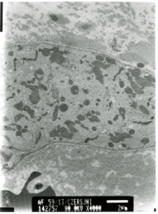
Electron microscopy analysis of the heart biopsy
Abstract:
Ultrastructural analysis revealed numerous changes in the analyzed biopsy. Within the cardiomyocytes large areas completely devoid of myofibrils were observed. Myofibrils disorganization was common (Figs. 1,2). In areas devoid of myofibrils, lipofuscin deposits were visible, as well as accumulation of altered mitochondria, characterized by a bright matrix and loss of mitochondrial cristae (Figs. 3-6). Features of laminopathy were observed. Nuclei of cardiomyocytes were deformed, with changes in the shape including numerous indentations inthe nuclear envelope. Many nuclei showed breaks in the nuclear membrane, which was associated with penetration of cell organelles into the nuclei (Figs. 7-12). Abundant fibrosis was observed in the extracellular space (Fig. 13).
Detailed Resource Type:
Format:
Resource Identifier:
Language:
Rights:
Creative Commons Attribution BY 4.0 license
Terms of use:
Copyright-protected material. [CC BY 4.0] May be used within the scope specified in Creative Commons Attribution BY 4.0 license, full text available at: ; -
Digitizing institution:
Mossakowski Medical Research Institute PAS
Original in:
Library of the Mossakowski Medical Research Institute PAS
Projects co-financed by:
Access:
Object collections:
- Digital Repository of Scientific Institutes > Partners' collections > Mossakowski Medical Research Institute PAS > Research data > File of heart diseases by Prof. A. Fidziańska-Dolot - ultrastructural studies
- Digital Repository of Scientific Institutes > Scientific data and objects > Medical science > File of medical cases
Last modified:
Feb 1, 2022
In our library since:
Feb 9, 2021
Number of object content downloads / hits:
55
All available object's versions:
https://rcin.org.pl./publication/194059
Show description in RDF format:
Show description in RDFa format:
Show description in OAI-PMH format:
| Edition name | Date |
|---|---|
| kardiomiopatia restrykcyjna - 56/13 | Feb 1, 2022 |
Objects Similar
Fidziańska-Dolot, Anna (1930–2015)
Fidziańska-Dolot, Anna (1930–2015)
Studies of heart ultrastructure in various diseases by prof A. Fidziańska-Dolot: myocarditis - 51/08
Fidziańska-Dolot, Anna (1930–2015)
Fidziańska-Dolot, Anna (1930–2015)
Fidziańska-Dolot, Anna (1930–2015)
Fidziańska-Dolot, Anna (1930–2015)
Fidziańska-Dolot, Anna (1930–2015)


