- O repozytorium
- Kolekcje
- Indeksy
- Historia przeglądania
-
Repozytoria RCIN
-
 INSTYTUT ARCHEOLOGII I ETNOLOGII POLSKIEJ AKADEMII NAUK
-
 INSTYTUT BADAŃ LITERACKICH POLSKIEJ AKADEMII NAUK
-
 INSTYTUT BADAWCZY LEŚNICTWA
-
 INSTYTUT BIOLOGII DOŚWIADCZALNEJ IM. MARCELEGO NENCKIEGO POLSKIEJ AKADEMII NAUK
-
 INSTYTUT BIOLOGII SSAKÓW POLSKIEJ AKADEMII NAUK
-
 INSTYTUT CHEMII FIZYCZNEJ PAN
-
 INSTYTUT CHEMII ORGANICZNEJ PAN
-
 INSTYTUT FILOZOFII I SOCJOLOGII PAN
-
 INSTYTUT GEOGRAFII I PRZESTRZENNEGO ZAGOSPODAROWANIA PAN
-
 INSTYTUT HISTORII im. TADEUSZA MANTEUFFLA POLSKIEJ AKADEMII NAUK
-
 INSTYTUT JĘZYKA POLSKIEGO POLSKIEJ AKADEMII NAUK
-
 INSTYTUT MATEMATYCZNY PAN
-
 INSTYTUT MEDYCYNY DOŚWIADCZALNEJ I KLINICZNEJ IM.MIROSŁAWA MOSSAKOWSKIEGO POLSKIEJ AKADEMII NAUK
-
 INSTYTUT PODSTAWOWYCH PROBLEMÓW TECHNIKI PAN
-
 INSTYTUT SLAWISTYKI PAN
-
 SIEĆ BADAWCZA ŁUKASIEWICZ - INSTYTUT TECHNOLOGII MATERIAŁÓW ELEKTRONICZNYCH
-
 MUZEUM I INSTYTUT ZOOLOGII POLSKIEJ AKADEMII NAUK
-
 INSTYTUT BADAŃ SYSTEMOWYCH PAN
-
 INSTYTUT BOTANIKI IM. WŁADYSŁAWA SZAFERA POLSKIEJ AKADEMII NAUK
-
- Wyszukaj w całym Repozytorium
- Piśmiennictwo i mapy
- Archeologia
- Baza Młynów
- Nauki przyrodnicze
Wyszukiwanie zaawansowane
Wyszukiwanie zaawansowane
Wyszukiwanie zaawansowane
Wyszukiwanie zaawansowane
Wyszukiwanie zaawansowane

Obiekt
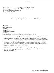
Tytuł: Kartoteka przypadków klinicznych chorób nerwowo-mięśniowych (2012) - opis 70/12
Współtwórca:
Miejsce wydania:
Opis:
Badanie wycinka mięśniowego w mikroskopie elektronowym
Abstrakt:
Rozpoznanie: Podejrzenie dystrofii mięśniowej. Miopatia. Fig. 1,2,3,4,5,6. Jądra komórkowe w znacznej większości włókien mięśniowych położone ośrodkowo, zwykle pojedyncze lub w liczbie dwóch. Fig 7.W niektórych włóknach na przekroju podłużnym widoczny ułożony ośrodkowo rządwielu jąder komórkowych, wyraźny jest ubytek miofibrylli. Fig. 8,9,10. Obserwuje się rozpad aparatu kurczliwego, nasilony zwłaszcza w pobliżu jąder komórek. Fig. 11. Występuje bardzo obfita tkanka łączna.
Szczegółowy typ zasobu:
Dokument mieszany ; Fotografia naukowa
Format:
Identyfikator zasobu:
Język:
Prawa:
Licencja Creative Commons Uznanie autorstwa 4.0
Zasady wykorzystania:
Zasób chroniony prawem autorskim. [CC BY 4.0 Międzynarodowe] Korzystanie dozwolone zgodnie z licencją Creative Commons Uznanie autorstwa 4.0, której pełne postanowienia dostępne są pod adresem: ; -
Digitalizacja:
Instytut Medycyny Doświadczalnej i Klinicznej im. M. Mossakowskiego Polskiej Akademii Nauk
Lokalizacja oryginału:
Biblioteka Instytutu Medycyny Doświadczalnej i Klinicznej im. M. Mossakowskiego PAN
Dofinansowane ze środków:
Dostęp:
Kolekcje, do których przypisany jest obiekt:
- Repozytorium Cyfrowe Instytutów Naukowych > Kolekcje Partnerów > Instytut Medycyny Doświadczalnej i Klinicznej PAN > Dane Badawcze > Kartoteka chorób nerwowo-mięśniowych Prof. A. Fidziańskiej-Dolot
- Repozytorium Cyfrowe Instytutów Naukowych > Dane i obiekty naukowe > Nauki medyczne > Kartoteka przypadków medycznych
Data ostatniej modyfikacji:
1 lut 2022
Data dodania obiektu:
21 kwi 2020
Liczba pobrań / odtworzeń:
34
Wszystkie dostępne wersje tego obiektu:
https://rcin.org.pl./publication/151012
Wyświetl opis w formacie RDF:
Wyświetl opis w formacie RDFa:
Wyświetl opis w formacie OAI-PMH:
| Nazwa wydania | Data |
|---|---|
| opis 70/12 | 1 lut 2022 |
Obiekty Podobne
Fidziańska-Dolot, Anna (1930–2015)
Fidziańska-Dolot, Anna (1930–2015)
Fidziańska-Dolot, Anna (1930–2015)
Fidziańska-Dolot, Anna (1930–2015)
Fidziańska-Dolot, Anna (1930–2015)
Fidziańska-Dolot, Anna (1930–2015)
Fidziańska-Dolot, Anna (1930–2015)
Fidziańska-Dolot, Anna (1930–2015)


