- About project
- Collections
- Indexes
- Recently viewed
-
RCIN Repositories
-
 INSTYTUT ARCHEOLOGII I ETNOLOGII POLSKIEJ AKADEMII NAUK
-
 INSTYTUT BADAŃ LITERACKICH POLSKIEJ AKADEMII NAUK
-
 INSTYTUT BADAWCZY LEŚNICTWA
-
 INSTYTUT BIOLOGII DOŚWIADCZALNEJ IM. MARCELEGO NENCKIEGO POLSKIEJ AKADEMII NAUK
-
 INSTYTUT BIOLOGII SSAKÓW POLSKIEJ AKADEMII NAUK
-
 INSTYTUT CHEMII FIZYCZNEJ PAN
-
 INSTYTUT CHEMII ORGANICZNEJ PAN
-
 INSTYTUT FILOZOFII I SOCJOLOGII PAN
-
 INSTYTUT GEOGRAFII I PRZESTRZENNEGO ZAGOSPODAROWANIA PAN
-
 INSTYTUT HISTORII im. TADEUSZA MANTEUFFLA POLSKIEJ AKADEMII NAUK
-
 INSTYTUT JĘZYKA POLSKIEGO POLSKIEJ AKADEMII NAUK
-
 INSTYTUT MATEMATYCZNY PAN
-
 INSTYTUT MEDYCYNY DOŚWIADCZALNEJ I KLINICZNEJ IM.MIROSŁAWA MOSSAKOWSKIEGO POLSKIEJ AKADEMII NAUK
-
 INSTYTUT PODSTAWOWYCH PROBLEMÓW TECHNIKI PAN
-
 INSTYTUT SLAWISTYKI PAN
-
 SIEĆ BADAWCZA ŁUKASIEWICZ - INSTYTUT TECHNOLOGII MATERIAŁÓW ELEKTRONICZNYCH
-
 MUZEUM I INSTYTUT ZOOLOGII POLSKIEJ AKADEMII NAUK
-
 INSTYTUT BADAŃ SYSTEMOWYCH PAN
-
 INSTYTUT BOTANIKI IM. WŁADYSŁAWA SZAFERA POLSKIEJ AKADEMII NAUK
-
- Search in all Repository
- Literature and maps
- Archeology
- Mills database
- Natural sciences
Advanced search
Advanced search
Advanced search
Advanced search
Advanced search

Object
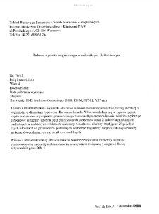
Title: Files for neuromuscular diseases (2012) - nr 70/12
Contributor:
Mossakowski Medical Research Center Polish Academy of Sciences. Department of Neuromuscular
Place of publishing:
Description:
Muscle segment examination in electron microscopy
Abstract:
Po A 4-year-old patient was examined. A biopsy of quadriceps sinister was performed. Electronmicroscopy analysis revealed ultrastructurally changed myofibres. The vast majority of muscle fibers were characterized by centrally located nuclei, usually single or in number of two (Fig. 1,2,3,4,5,6.) In some fibers in the longitudinal section we observed a centrally arranged row of many nuclei and the evident loss of myofibril was seen (Fig. 7). The disruption of the contractile apparatus was observed, especially near the cell nuclei (Fig. 8,9,10). Abundant connective tissue was visible (Fig. 11). Ultrastructural and clinical picture suggests centronuclear myopathy associated with inappropriate BIN1 gene activity.
Detailed Resource Type:
Format:
Resource Identifier:
Language:
Rights:
Creative Commons Attribution BY 4.0 license
Terms of use:
Copyright-protected material. [CC BY 4.0] May be used within the scope specified in Creative Commons Attribution BY 4.0 license, full text available at: ; -
Digitizing institution:
Mossakowski Medical Research Institute PAS
Original in:
Library of the Mossakowski Medical Research Institute PAS
Projects co-financed by:
Access:
Object collections:
- Digital Repository of Scientific Institutes > Partners' collections > Mossakowski Medical Research Institute PAS > Research data > File of neuromuscular diseases of Prof. A. Fidziańska-Dolot
- Digital Repository of Scientific Institutes > Scientific data and objects > Medical science > File of medical cases
Last modified:
Feb 1, 2022
In our library since:
Apr 21, 2020
Number of object content downloads / hits:
34
All available object's versions:
https://rcin.org.pl./publication/151012
Show description in RDF format:
Show description in RDFa format:
Show description in OAI-PMH format:
| Edition name | Date |
|---|---|
| opis 70/12 | Feb 1, 2022 |
Objects Similar
Fidziańska-Dolot, Anna (1930–2015)
Fidziańska-Dolot, Anna (1930–2015)
Fidziańska-Dolot, Anna (1930–2015)
Fidziańska-Dolot, Anna (1930–2015)


