- Search in all Repository
- Literature and maps
- Archeology
- Mills database
- Natural sciences
Advanced search
Advanced search
Advanced search
Advanced search
Advanced search

Object
Title: Files for neuromuscular diseases (2012) - nr 61/12
Contributor:
Mossakowski Medical Research Center Polish Academy of Sciences. Department of Neuromuscular
Place of publishing:
Description:
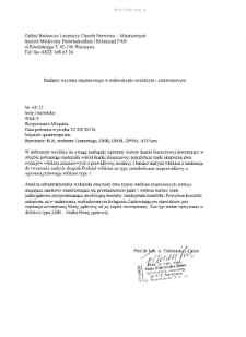
Badanie wycinka mięśniowego w mikroskopie świetlnym i elektronowym
Abstract:
A 9-year-old patient was examined. A biopsy of quadriceps sinister was performed. Electronmicroscopy analysis revealed two types of fibers. First type was characterized by disruption the contractile apparatus and the accumulation of nuclei. Abundant fibrous tissue, especially collagen was observed (Fig. 1,2). Second type of fibers were small diameter, had one nucleus and reminiscent myotube morphology (Fig. 3). In this type of fibers separation of the outer nuclear membrane from its inner part were observed (Fig. 4,5).
Detailed Resource Type:
Format:
Resource Identifier:
Language:
Rights:
Creative Commons Attribution BY 4.0 license
Terms of use:
Copyright-protected material. [CC BY 4.0] May be used within the scope specified in Creative Commons Attribution BY 4.0 license, full text available at: ; -
Digitizing institution:
Mossakowski Medical Research Institute PAS
Original in:
Library of the Mossakowski Medical Research Institute PAS
Projects co-financed by:
Access:
Object collections:
- Digital Repository of Scientific Institutes > Partners' collections > Mossakowski Medical Research Institute PAS > Research data > File of neuromuscular diseases of Prof. A. Fidziańska-Dolot
- Digital Repository of Scientific Institutes > Scientific data and objects > Medical science > File of medical cases
Last modified:
Feb 1, 2022
In our library since:
Apr 20, 2020
Number of object content downloads / hits:
45
All available object's versions:
https://rcin.org.pl./publication/112268
Show description in RDF format:
Show description in RDFa format:
Show description in OAI-PMH format:
| Edition name | Date |
|---|---|
| opis 61/12 | Feb 1, 2022 |

 INSTYTUT ARCHEOLOGII I ETNOLOGII POLSKIEJ AKADEMII NAUK
 INSTYTUT BADAŃ LITERACKICH POLSKIEJ AKADEMII NAUK
 INSTYTUT BADAWCZY LEŚNICTWA
 INSTYTUT BIOLOGII DOŚWIADCZALNEJ IM. MARCELEGO NENCKIEGO POLSKIEJ AKADEMII NAUK
 INSTYTUT BIOLOGII SSAKÓW POLSKIEJ AKADEMII NAUK
 INSTYTUT CHEMII FIZYCZNEJ PAN
 INSTYTUT CHEMII ORGANICZNEJ PAN
 INSTYTUT FILOZOFII I SOCJOLOGII PAN
 INSTYTUT GEOGRAFII I PRZESTRZENNEGO ZAGOSPODAROWANIA PAN
 INSTYTUT HISTORII im. TADEUSZA MANTEUFFLA POLSKIEJ AKADEMII NAUK
 INSTYTUT JĘZYKA POLSKIEGO POLSKIEJ AKADEMII NAUK
 INSTYTUT MATEMATYCZNY PAN
 INSTYTUT MEDYCYNY DOŚWIADCZALNEJ I KLINICZNEJ IM.MIROSŁAWA MOSSAKOWSKIEGO POLSKIEJ AKADEMII NAUK
 INSTYTUT PODSTAWOWYCH PROBLEMÓW TECHNIKI PAN
 INSTYTUT SLAWISTYKI PAN
 SIEĆ BADAWCZA ŁUKASIEWICZ - INSTYTUT TECHNOLOGII MATERIAŁÓW ELEKTRONICZNYCH
 MUZEUM I INSTYTUT ZOOLOGII POLSKIEJ AKADEMII NAUK
 INSTYTUT BADAŃ SYSTEMOWYCH PAN
 INSTYTUT BOTANIKI IM. WŁADYSŁAWA SZAFERA POLSKIEJ AKADEMII NAUK


