- Search in all Repository
- Literature and maps
- Archeology
- Mills database
- Natural sciences
Advanced search
Advanced search
Advanced search
Advanced search
Advanced search

Object
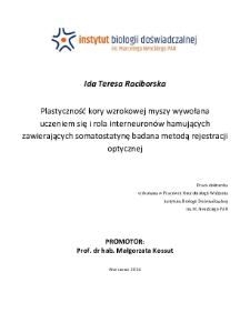
Title: Plastyczność kory wzrokowej myszy wywołana uczeniem się i rola interneuronów hamujących zawierających somatostatynę badana metodą rejestracji optycznej : praca doktorska
Institutional creator:
Instytut Biologii Doświadczalnej im. Marcelego Nenckiego PAN
Contributor:
Kossut, Małgorzata : Supervisor
Publisher:
Instytut Biologii Doświadczalnej im. Marcelego Nenckiego PAN
Place of publishing:
Description:
251 pages : illustrations ; 30 cm ; Bibliography ; Summary in English
Degree name:
Degree discipline :
Degree grantor:
Nencki Institute of Experimental Biology PAS ; degree obtained in 2024
Type of object:
Abstract:
Plasticity is a key feature of virtually all brains. It is hypothesized to underpin learning and memory and plays an important role in the early development of brain circuits. However, plasticity is not limited to developmental phases. This capacity is preserved in adulthood in the mammalian visual cortex and other basic sensory centers. Partial retinal lesions are a well-established model for inducing adult plasticity in the visual cortex, but our knowledge about how the inhibitory network contributes to brain plasticity remains incomplete. Somatostatin (SST-IN) interneurons constitute a significant neocortical subpopulation of interneurons, along with parvalbumin (PV-IN) and vasoactive intestinal peptide (VIP- IN) interneurons. Unlike the extensively studied PV-interneurons, which are acknowledged as key components in guiding ocular dominance plasticity, the contribution of SST-interneurons is less understood because neuroplasticity has only been more intensively studied in the last decade. In this dissertation, a fear conditioning mouse model was used to investigate plasticity in adults by recording the activity of the entire primary visual cortex (V1), with a particular emphasis on the role of plastic changes induced by learning through different interneurons expressing SST-IN and PV-IN. In the first stage of this study, an intrinsic signal optical imaging (ISOI) technique was used to investigate the stimulus orientation sensitivities of neurons in the mouse visual cortex in vivo. Optical signals from the right and left V1 cortex were recorded in response to moving black and white bars. ISOI allowed cortical activity to be recorded in the form of activation maps and optical signals. The second stage was devoted to investigating the effects of classical conditioning, in which orientation- specific visual stimuli were coupled to caudal current application (UCS). Animals were divided into experimental groups: those subjected to full classical conditioning, pseudoclassical conditioning, and exposure only to visual stimuli. The learning indicator was the induction of conditioned bradycardia. Training sessions lasted 10 minutes each for seven consecutive days. ECG was monitored during the training. Twenty-four hours after training, its effect on visual cortex V1 activity was recorded using the ISOI technique. It was found that conditioning led to increased activation strength in the visual cortex compared to the control group. Additionally, assessment of the active areas' size revealed a concentration of activity. The third step was to investigate the importance of SST-IN in the process of plastic changes induced by conditioning and their involvement in the activation of visual cortex V1. It was investigated whether the experimental procedures used affected the abundance of SST-IN and PV-IN in different areas of the visual cortex, in all its layers. After finding changes in SST-IN abundance in the conditioned group, it was tested whether chemogenetic silencing of SST-IN would affect the outcome of conditioning. SST interneurons appear to play a key role in the occurrence of plastic conditioning-induced changes, as the experimental group in which SST-IN activity was blocked using DREADD did not show changes induced by classical conditioning. These findings affirm the utility of ISOI as a technique for imaging the activity of large neuron populations in the mouse visual cortex, allowing for the characterization of orientation sensitivity, as well as changes in light scattering response amplitudes during ISOI registration. The study demonstrated that SST-IN activity in the cortex is essential for forming associations between conditioned and unconditioned stimuli, confirming its crucial role in shaping neuroplastic changes related to conditioning.
Detailed Resource Type:
Resource Identifier:
Source:
Language:
Language of abstract:
Rights:
Terms of use:
Copyright-protected material. May be used within the limits of statutory user freedoms
Copyright holder:
Publication made available with the written permission of the author
Digitizing institution:
Nencki Institute of Experimental Biology of the Polish Academy of Sciences
Original in:
Library of the Nencki Institute of Experimental Biology PAS
Access:
Object collections:
- Digital Repository of Scientific Institutes > Partners' collections > Nencki Institute of Experimental Biology PAS
- Digital Repository of Scientific Institutes > Partners' collections > Nencki Institute of Experimental Biology PAS > Dissertations
- Digital Repository of Scientific Institutes > Partners' collections > Nencki Institute of Experimental Biology PAS > Dissertations > PhD Thesis
- Digital Repository of Scientific Institutes > Literature
- Digital Repository of Scientific Institutes > Literature > Thesis
Last modified:
Dec 12, 2024
In our library since:
Oct 28, 2024
Number of object content downloads / hits:
2
All available object's versions:
https://rcin.org.pl./publication/279277
Show description in RDF format:
Show description in RDFa format:
Show description in OAI-PMH format:
Objects Similar
Kanigowski, Dominik
Grabowska, Agnieszka Kamila
Dzięgiel-Fivet, Gabriela

 INSTYTUT ARCHEOLOGII I ETNOLOGII POLSKIEJ AKADEMII NAUK
 INSTYTUT BADAŃ LITERACKICH POLSKIEJ AKADEMII NAUK
 INSTYTUT BADAWCZY LEŚNICTWA
 INSTYTUT BIOLOGII DOŚWIADCZALNEJ IM. MARCELEGO NENCKIEGO POLSKIEJ AKADEMII NAUK
 INSTYTUT BIOLOGII SSAKÓW POLSKIEJ AKADEMII NAUK
 INSTYTUT CHEMII FIZYCZNEJ PAN
 INSTYTUT CHEMII ORGANICZNEJ PAN
 INSTYTUT FILOZOFII I SOCJOLOGII PAN
 INSTYTUT GEOGRAFII I PRZESTRZENNEGO ZAGOSPODAROWANIA PAN
 INSTYTUT HISTORII im. TADEUSZA MANTEUFFLA POLSKIEJ AKADEMII NAUK
 INSTYTUT JĘZYKA POLSKIEGO POLSKIEJ AKADEMII NAUK
 INSTYTUT MATEMATYCZNY PAN
 INSTYTUT MEDYCYNY DOŚWIADCZALNEJ I KLINICZNEJ IM.MIROSŁAWA MOSSAKOWSKIEGO POLSKIEJ AKADEMII NAUK
 INSTYTUT PODSTAWOWYCH PROBLEMÓW TECHNIKI PAN
 INSTYTUT SLAWISTYKI PAN
 SIEĆ BADAWCZA ŁUKASIEWICZ - INSTYTUT TECHNOLOGII MATERIAŁÓW ELEKTRONICZNYCH
 MUZEUM I INSTYTUT ZOOLOGII POLSKIEJ AKADEMII NAUK
 INSTYTUT BADAŃ SYSTEMOWYCH PAN
 INSTYTUT BOTANIKI IM. WŁADYSŁAWA SZAFERA POLSKIEJ AKADEMII NAUK


