- Search in all Repository
- Literature and maps
- Archeology
- Mills database
- Natural sciences
Advanced search
Advanced search
Advanced search
Advanced search
Advanced search

Object
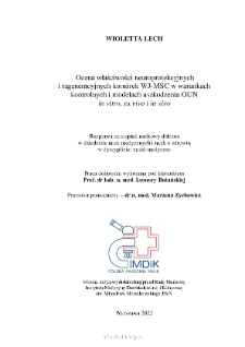
Title: Ocena właściwości neuroprotekcyjnych i regeneracyjnych komórek WJ-MSC w warunkach uszkodzenia OUN in vitro, ex vivo i in vivo
Institutional creator:
Instytut Medycyny Doświadczalnej i Klinicznej im. M. Mossakowskiego PAN
Contributor:
Bużańska, Leonora (Promotor) ; Zychowicz, Marzena (Promotor pomocniczy)
Place of publishing:
Description:
Bibliografia od str. 187-201 ; 201 s.: il., wykr., ryc., fotogr.; 30 cm
Degree name:
Level of degree:
Degree discipline :
Abstract:
Cell therapy is a branch of regenerative medicine and relates to cell transplantations in order to rebuild damaged tissue/organ when pharmacological and surgical treatment is insufficient or ineffective. Stem cells characterized by potential to self-renew and multilineage differentiation are most commonly used for transplantations in regenerative medicine. Ischemic stroke was selected as a model of CNS damage to analyze the influence of the microenvironment and preconditioning in the pre-transplantation phase on the stem cells isolated from Wharton’s Jelly (WJ-MSCs) pro-regenerative properties.The endogenous stem cells microenvironment, known as the “niche”, determines their survival and growth, and enables further differentiation. Therefore, an important element of this work was to optimize the biomimetic microenvironment in vitro, which mimics the conditions similar to the endogenous: 1) control – WJ-MSCs cultured in 3D conditions in a hydrogels (made from platelet lysate or fibrinogen) resembling the composition and mechanical properties of nervous tissue and under 5% oxygen concentration, typical of the stem cell niche in the brain; 2) pathological – additional stimulation under the conditions as above with the pro-inflammatory factors.It was shown that the 3D hydrogel scaffolds create a structure that allows the cells deposition, their high survival, and migration beyond the scaffolds. Moreover, an increased expression of BDNF, GDNF, VEGF-A, TGF-β1, IL-6, IL-1β were observed under both 21% and 5% O2. Additionally, in WJ-MSCs cultured in 3D scaffolds, an increased expression of neural markers (nestin, β-Tubulin III, NF-200, GFAP) was observed at the mRNA and protein levels. The 3D culture conditions significantly increased the response of WJ-MSCs to the pro-inflammatory factors – a significantly increased mRNA expression of trophic factors – BDNF, GDNF, VEGF-A, and immunomodulatory factors – TGF-β1, IL-6, IL-1β was observed.In ex vivo studies, the OGD model (temporary oxygen and glucose deprivation) was used, which imitates an ischemic injury. The strongest effect was observed after the co-culture with WJ-MSCs preincubated in 5% O2 and fibrin scaffolds. An increased expression of GDNF, VEGF-A and a decreased expression of pro-inflammatory IL-1β with a simultaneous increase in the expression of the anti-inflammatory TGF-β1 was observed in WJ-MSCs encapsulated in scaffolds and co-cultured with damaged organotypic hippocampal slices.In vivo studies were conducted using an experimental model of rat brain injury. The signal from WJ-MSCs labelled with iron oxide nanoparticles was observed in the injection site (striatum) 24 hours after transplantation, and also 7-, 14-, and 21- days post-transplantation. In addition, after the WJ-MSCs transplantation in 2D or 3D, a signal was detected at the lesionsite during diffusion-weighted imaging (DWI), which was not observed in sham groups or after only the focal injury.After the WJ-MSCs transplantation, a decreased size of the brain-damaged area and an increased expression of rat markers: BDNF, GDNF, VEGF-A, TGF-β1 were detected. The highest increase in the genes’ expression level was observed after transplantation of the cells that were cultured in physiological normoxia conditions (5% O2) and then transplanted in scaffolds. Transplantation of WJ-MSCs caused also the decreased expression of rat IL-6 and IL-1β, especially after 24 hours.The obtained results showed that the hydrogel scaffolds made of platelet lysate and fibrinogen used in this study are promising material for potential use in cell therapy of central nervous system disorders. The most preferred therapeutic approach is the maintaining of the cells in culture under physiological normoxia (5% O2) followed by their transplantation as encapsulated in hydrogel scaffolds. These scaffolds should maintain mechanical properties similar to those found in the brain.
Detailed Resource Type:
Format:
Resource Identifier:
Source:
Language:
Rights:
Creative Commons Attribution BY 4.0 license
Terms of use:
Copyright-protected material. [CC BY 4.0] May be used within the scope specified in Creative Commons Attribution BY 4.0 license, full text available at: ; -
Digitizing institution:
Mossakowski Medical Research Institute PAS
Original in:
Library of the Mossakowski Medical Research Institute PAS
Projects co-financed by:
Access:
Object collections:
- Digital Repository of Scientific Institutes > Partners' collections > Mossakowski Medical Research Institute PAS
- Digital Repository of Scientific Institutes > Partners' collections > Mossakowski Medical Research Institute PAS > Theses > Ph.D Dissertationes
Last modified:
Mar 1, 2024
In our library since:
Feb 28, 2024
Number of object content downloads / hits:
113
All available object's versions:
https://rcin.org.pl./publication/273568
Show description in RDF format:
Show description in RDFa format:
Show description in OAI-PMH format:
Objects Similar
Gannushkina I.V. Mossakowski, Mirosław Jan (1929 - 2001) i in.
Niebrój-Dobosz, Irena Rafałowska, Janina Łukasiuk, Mirosława Pfeffer, Anna Mossakowski, Mirosław Jan (1929–2001)
Doleżyczek, Hubert
Skowrońska, Katarzyna
Łazarewicz, Jerzy W. Salińska, Elżbieta

 INSTYTUT ARCHEOLOGII I ETNOLOGII POLSKIEJ AKADEMII NAUK
 INSTYTUT BADAŃ LITERACKICH POLSKIEJ AKADEMII NAUK
 INSTYTUT BADAWCZY LEŚNICTWA
 INSTYTUT BIOLOGII DOŚWIADCZALNEJ IM. MARCELEGO NENCKIEGO POLSKIEJ AKADEMII NAUK
 INSTYTUT BIOLOGII SSAKÓW POLSKIEJ AKADEMII NAUK
 INSTYTUT CHEMII FIZYCZNEJ PAN
 INSTYTUT CHEMII ORGANICZNEJ PAN
 INSTYTUT FILOZOFII I SOCJOLOGII PAN
 INSTYTUT GEOGRAFII I PRZESTRZENNEGO ZAGOSPODAROWANIA PAN
 INSTYTUT HISTORII im. TADEUSZA MANTEUFFLA POLSKIEJ AKADEMII NAUK
 INSTYTUT JĘZYKA POLSKIEGO POLSKIEJ AKADEMII NAUK
 INSTYTUT MATEMATYCZNY PAN
 INSTYTUT MEDYCYNY DOŚWIADCZALNEJ I KLINICZNEJ IM.MIROSŁAWA MOSSAKOWSKIEGO POLSKIEJ AKADEMII NAUK
 INSTYTUT PODSTAWOWYCH PROBLEMÓW TECHNIKI PAN
 INSTYTUT SLAWISTYKI PAN
 SIEĆ BADAWCZA ŁUKASIEWICZ - INSTYTUT TECHNOLOGII MATERIAŁÓW ELEKTRONICZNYCH
 MUZEUM I INSTYTUT ZOOLOGII POLSKIEJ AKADEMII NAUK
 INSTYTUT BADAŃ SYSTEMOWYCH PAN
 INSTYTUT BOTANIKI IM. WŁADYSŁAWA SZAFERA POLSKIEJ AKADEMII NAUK


