- Search in all Repository
- Literature and maps
- Archeology
- Mills database
- Natural sciences
Advanced search
Advanced search
Advanced search
Advanced search
Advanced search

Object
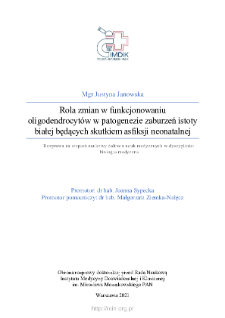
Title: Rola zmian w funkcjonowaniu oligodendrocytów w patogenezie zaburzeń istoty białej będących skutkiem asfiksji neonatalnej
Contributor:
Sypecka, Joanna (Promotor) ; Ziemka-Nałęcz, Małgorzata (Promotor pomocniczy)
Publisher:
Instytut Medycyny Doświadczalnej i Klincznej im. M. Mossakowskiego PAN
Place of publishing:
Degree name:
Level of degree:
Degree discipline :
Degree grantor:
Instytut Medycyny Doświadczalnej i Klinicznej im. M. Mossakowskiego PAN
Type of object:
Abstract:
Perinatal asphyxia (otherwise known as hypoxia-ischemia, HI) is the second most common cause of neonatal death worldwide. Causes of asphyxia include premature birth and often result in disruption of normal brain development, leading to hypoxic-ischemic encephalopathy (HIE). In children with mild asphyxia further development is usually normal, but severe hypoxia contributes to the development of infantile cerebral palsy, epilepsy, and may increase the risk of developing autism spectrum disorders. Diffuse damage to white matter, that is oligodendrocytes and their myelin sheaths, are among the most frequently detected pathological changes in HIE. Myelin is an electrical insulator that conditions fast propagation of nerve impulses and forms an indispensable protective barrier for axons. In the developing brain oligodendrocytes differentiate into so-called oligodendrocyte progenitor cells (OPCs). The OPCs differentiation into mature cells is most intense in the perinatal period and is regulated by, among others, trophic factors. Disturbance of neural tissue homeostasis induced by neonatal asphyxia may therefore influence the differentiation and further functioning of oligodendrocytes. Previous studies have shown that OPCs may provide neuroprotective factors, particularly in the region of damaged neural tissue. In the present study, we hypothesized that after HI, OPCs are characterized by an impaired growth and differentiation protocol that promotes their maintenance in a progenitor state and limits maturation. In this study, the potential of OPCs to secrete a selected neurotrophic factor, IGF-1, was evaluated. In addition, the changes in IGF-1 levels in neural tissue after HI injury and the potential of this factor as a therapy for HIE were investigated. Studies were performed using three HI models: in vivo (cerebral hypoxia-ischemia in 7-day-old rats); ex vivo (organotypic cultures of hippocampal slices subjected to temporary nutrient and oxygen deprivation - OGD) and in vitro (cultures of primary rat neonatal OPCs subjected to OGD). The results obtained show that in the first days after damage, OPCs do not show reduced viability but intensively proliferate, and myelin protein expression increases simultaneously. However, analysis of myelination in adult rats (10 weeks after HI) confirmed the typical clinical pattern of neonatal hypoxia-ischemia, i.e., hypotrophy of the hippocampus, corpus callosum, and striatum, as well as areas of hypomyelination in the cortex, striatum, and CA3 region and also delamination of the myelin sheets. Analysis of IGF-1 levels in neural tissue in an in vivo model showed a significant increase in IGF-1 levels after HI on the first day after injury. Ex vivo and in vitro studies showed that OPCs can be a source of IGF-1 and that OGD decreases its secretion. Supplementation of the culture medium with IGF-1 at a concentration of 50 ng/ml induced an increase in the rate of cell proliferation, while a low concentration of 10 ng/ml IGF-1 promoted the production of cell branches, which may imply the promotion of maturation of these cells. The results of the experiments conducted in the present study showed that periodic increases in IGF-1 levels, among others, may contribute to the changes in OPCs differentiation. Despite the increase in myelin protein expression after HI, the production of functional myelin sheaths is impaired. Our study indicates that IGF-1 is one of the factors that may modulate the ability of oligodendrocytes to myelinate after asphyxia, and may be considered as a potential drug to promote white matter development.
Detailed Resource Type:
Resource Identifier:
Source:
IMDiK PAN, sygn. ZS409 ; click here to follow the link
Language:
Language of abstract:
Rights:
Creative Commons Attribution BY 4.0 license
Terms of use:
Copyright-protected material. [CC BY 4.0] May be used within the scope specified in Creative Commons Attribution BY 4.0 license, full text available at: ; -
Digitizing institution:
Mossakowski Medical Research Institute PAS
Original in:
Library of the Mossakowski Medical Research Institute PAS
Access:
Object collections:
- Digital Repository of Scientific Institutes > Partners' collections > Mossakowski Medical Research Institute PAS > Theses
- Digital Repository of Scientific Institutes > Partners' collections > Mossakowski Medical Research Institute PAS > Theses > Ph.D Dissertationes
Last modified:
Nov 21, 2024
In our library since:
May 12, 2022
Number of object content downloads / hits:
78
All available object's versions:
https://rcin.org.pl./publication/272055

 INSTYTUT ARCHEOLOGII I ETNOLOGII POLSKIEJ AKADEMII NAUK
 INSTYTUT BADAŃ LITERACKICH POLSKIEJ AKADEMII NAUK
 INSTYTUT BADAWCZY LEŚNICTWA
 INSTYTUT BIOLOGII DOŚWIADCZALNEJ IM. MARCELEGO NENCKIEGO POLSKIEJ AKADEMII NAUK
 INSTYTUT BIOLOGII SSAKÓW POLSKIEJ AKADEMII NAUK
 INSTYTUT CHEMII FIZYCZNEJ PAN
 INSTYTUT CHEMII ORGANICZNEJ PAN
 INSTYTUT FILOZOFII I SOCJOLOGII PAN
 INSTYTUT GEOGRAFII I PRZESTRZENNEGO ZAGOSPODAROWANIA PAN
 INSTYTUT HISTORII im. TADEUSZA MANTEUFFLA POLSKIEJ AKADEMII NAUK
 INSTYTUT JĘZYKA POLSKIEGO POLSKIEJ AKADEMII NAUK
 INSTYTUT MATEMATYCZNY PAN
 INSTYTUT MEDYCYNY DOŚWIADCZALNEJ I KLINICZNEJ IM.MIROSŁAWA MOSSAKOWSKIEGO POLSKIEJ AKADEMII NAUK
 INSTYTUT PODSTAWOWYCH PROBLEMÓW TECHNIKI PAN
 INSTYTUT SLAWISTYKI PAN
 SIEĆ BADAWCZA ŁUKASIEWICZ - INSTYTUT TECHNOLOGII MATERIAŁÓW ELEKTRONICZNYCH
 MUZEUM I INSTYTUT ZOOLOGII POLSKIEJ AKADEMII NAUK
 INSTYTUT BADAŃ SYSTEMOWYCH PAN
 INSTYTUT BOTANIKI IM. WŁADYSŁAWA SZAFERA POLSKIEJ AKADEMII NAUK


