- Wyszukaj w całym Repozytorium
- Piśmiennictwo i mapy
- Archeologia
- Baza Młynów
- Nauki przyrodnicze
Wyszukiwanie zaawansowane
Wyszukiwanie zaawansowane
Wyszukiwanie zaawansowane
Wyszukiwanie zaawansowane
Wyszukiwanie zaawansowane

Obiekt
Tytuł: Kartoteka przypadków klinicznych chorób nerwowo-mięśniowych (2011) - opis nr 1/11-1
Współtwórca:
Miejsce wydania:
Opis:
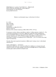
Badanie wycinka mięśniowego w mikroskopie świetlnym
Abstrakt:
Włókna mięśniowe o prawidłowej średnicy,zachowanej architekturze miofibryli i strukturze sarkomerów oraz włókna mniejsze,oddzielone dość obfitą tkanką łączną i tłuszczową. Występują jądra komórek mięśniowych o nieprawidłowj budowie, charakteryzujące się pofałdowanymi wypustkami. Obsewuje się zwiększoną ilość fropli tłuszczu.
Szczegółowy typ zasobu:
Dokument mieszany ; Fotografia naukowa
Format:
Identyfikator zasobu:
Język:
Prawa:
Licencja Creative Commons Uznanie autorstwa 4.0
Zasady wykorzystania:
Zasób chroniony prawem autorskim. [CC BY 4.0 Międzynarodowe] Korzystanie dozwolone zgodnie z licencją Creative Commons Uznanie autorstwa 4.0, której pełne postanowienia dostępne są pod adresem: ; -
Digitalizacja:
Instytut Medycyny Doświadczalnej i Klinicznej im. M. Mossakowskiego Polskiej Akademii Nauk
Lokalizacja oryginału:
Biblioteka Instytutu Medycyny Doświadczalnej i Klinicznej im. M. Mossakowskiego PAN
Dofinansowane ze środków:
Dostęp:
Kolekcje, do których przypisany jest obiekt:
- Repozytorium Cyfrowe Instytutów Naukowych > Kolekcje Partnerów > Instytut Medycyny Doświadczalnej i Klinicznej PAN > Dane Badawcze > Kartoteka chorób nerwowo-mięśniowych Prof. A. Fidziańskiej-Dolot
- Repozytorium Cyfrowe Instytutów Naukowych > Dane i obiekty naukowe > Nauki medyczne > Kartoteka przypadków medycznych
Data ostatniej modyfikacji:
1 lut 2022
Data dodania obiektu:
8 wrz 2020
Liczba pobrań / odtworzeń:
61
Wszystkie dostępne wersje tego obiektu:
https://rcin.org.pl./publication/110474
Wyświetl opis w formacie RDF:
Wyświetl opis w formacie RDFa:
Wyświetl opis w formacie OAI-PMH:
| Nazwa wydania | Data |
|---|---|
| opis nr 1/11 | 1 lut 2022 |
Obiekty Podobne
Fidziańska-Dolot, Anna (1930–2015)
Fidziańska-Dolot, Anna (1930–2015)
Fidziańska-Dolot, Anna (1930–2015)
Fidziańska-Dolot, Anna (1930–2015)
Fidziańska-Dolot, Anna (1930–2015)
Fidziańska-Dolot, Anna (1930–2015)
Fidziańska-Dolot, Anna (1930–2015)
Fidziańska-Dolot, Anna (1930–2015)

 INSTYTUT ARCHEOLOGII I ETNOLOGII POLSKIEJ AKADEMII NAUK
 INSTYTUT BADAŃ LITERACKICH POLSKIEJ AKADEMII NAUK
 INSTYTUT BADAWCZY LEŚNICTWA
 INSTYTUT BIOLOGII DOŚWIADCZALNEJ IM. MARCELEGO NENCKIEGO POLSKIEJ AKADEMII NAUK
 INSTYTUT BIOLOGII SSAKÓW POLSKIEJ AKADEMII NAUK
 INSTYTUT CHEMII FIZYCZNEJ PAN
 INSTYTUT CHEMII ORGANICZNEJ PAN
 INSTYTUT FILOZOFII I SOCJOLOGII PAN
 INSTYTUT GEOGRAFII I PRZESTRZENNEGO ZAGOSPODAROWANIA PAN
 INSTYTUT HISTORII im. TADEUSZA MANTEUFFLA POLSKIEJ AKADEMII NAUK
 INSTYTUT JĘZYKA POLSKIEGO POLSKIEJ AKADEMII NAUK
 INSTYTUT MATEMATYCZNY PAN
 INSTYTUT MEDYCYNY DOŚWIADCZALNEJ I KLINICZNEJ IM.MIROSŁAWA MOSSAKOWSKIEGO POLSKIEJ AKADEMII NAUK
 INSTYTUT PODSTAWOWYCH PROBLEMÓW TECHNIKI PAN
 INSTYTUT SLAWISTYKI PAN
 SIEĆ BADAWCZA ŁUKASIEWICZ - INSTYTUT TECHNOLOGII MATERIAŁÓW ELEKTRONICZNYCH
 MUZEUM I INSTYTUT ZOOLOGII POLSKIEJ AKADEMII NAUK
 INSTYTUT BADAŃ SYSTEMOWYCH PAN
 INSTYTUT BOTANIKI IM. WŁADYSŁAWA SZAFERA POLSKIEJ AKADEMII NAUK


